. 2012 Mar 22;3(3):e287.
doi: 10.1038/cddis.2012.23.

MicroRNA-34a upregulation during seizure-induced neuronal death

Affiliations

MicroRNA-34a upregulation during seizure-induced neuronal death

T Sano et al. Cell Death Dis. .

Abstract

MicroRNAs (miRNAs) are short, noncoding RNAs that function as posttranscriptional regulators of gene expression by controlling translation of mRNAs. A subset of miRNAs may be critical for the control of cell death, including the p53-regulated miRNA, miR-34a. Because seizures activate p53, and p53-deficient mice are reportedly resistant to damage caused by prolonged seizures, we investigated the role of miR-34a in seizure-induced neuronal death in vivo. Status epilepticus was induced by intra-amygdala microinjection of kainic acid in mice. This led to an early (2 h) multifold upregulation of miR-34a in the CA3 and CA1 hippocampal subfields and lower protein levels of mitogen-activated kinase kinase kinase 9, a validated miR-34a target. Immunoprecipitation of the RNA-induced silencing complex component, Argonaute-2, eluted significantly higher levels of miR-34a after seizures. Injection of mice with pifithrin-α, a putative p53 inhibitor, prevented miR-34a upregulation after seizures. Intracerebroventricular injection of antagomirs targeting miR-34a reduced hippocampal miR-34a levels and had a small modulatory effect on apoptosis-associated signaling, but did not prevent hippocampal neuronal death in models of either severe or moderate severity status epilepticus. Thus, prolonged seizures cause subfield-specific, temporally restricted upregulation of miR-34a, which may be p53 dependent, but miR-34a is probably not important for seizure-induced neuronal death in this model.


Figures
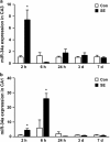
Figure 1
Figure 1
SE induces rapid upregulation of miR-34a. (a and b) RT-PCR measurement of mature miR-34a levels in CA3 and CA1 subfields from control (Con) mice and animals subjected to SE at various time points. Expression levels were corrected to RNU6B. *P<0.05 compared with control (n=4 per group)
Figure 2
Figure 2
RISC loading of miR-34a, downregulation of target Map3k9 and p53 dependence. (a) Graph showing miR-34a levels eluted by immunoprecipitation of Ago-2 from non-seizure controls (Con) and mice 2 h after SE. ***P<0.001 compared with control (n=3 per group). (b) Representative western blot (n=1 per lane) showing protein levels of the miR-34a target Map3k9 in Con and 2, 4 or 24 h after SE. β-Actin is included as a guide to protein loading. Molecular weight markers depicted to the right. Time in hours after KA is shown above. (c) Semiquantification of Map3k9 levels after seizures. *P<0.05 compared with control (n=4 per group). (d) RT-PCR measurement of p53-target gene p21WAF/Cip1 expression in seizure mice at 2 h showing reduced expression of the p53-regulated gene in mice treated with pifithrin-α (PFT) compared with vehicle (Veh, DMSO; *P<0.05 compared with vehicle; n=5 per group). (e) Graph showing RT-PCR measurement of miR-34a in animals given PFT or Veh before SE. **P<0.01 compared with vehicle (n=3–4 per group)
Figure 3
Figure 3
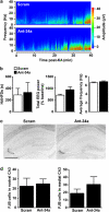
Effect of antagomirs on brain levels of miR-34a and seizure EEG. (a and b) Effect of antagomirs targeting miR-34a (Ant-34a) and scrambled (Scram) on hippocampal CA3 expression of A, miR-34a, and B, miR-134, measured 24 h after injection. Expression levels were corrected to RNU6B. *P<0.05 compared with Scram (n=3 per group). (c) Representative EEG spectrogram during SE in mice given either Scram or Ant-34a. (d) Graphs showing semiquantification of seizure parameters between treated mice subjected to SE (n=7–8 per group). Differences were not statistically significant
Figure 4
Figure 4
No effect of antagomirs targeting miR-34a on seizure-induced neuronal death. (a) Representative photomicrographs of the ipsilateral hippocampus at two levels of the dorsal hippocampus from mice 24 h after SE given either 0.5 nmol Scram or Ant-34a. Black dots are FJB-positive cells. Scale bars; rostral, 550 μm; medial, 600 μm. (b and c) Graphs showing FJB counts in ipsilateral CA3 and CA1 at rostral and medial levels of the dorsal hippocampus 24 h after SE (n=7–8 per group). Differences were not statistically significant
Figure 5
Figure 5
Antagomirs targeting miR-34a do not prevent neuronal death caused by moderate severity SE. (a) Representative seizure EEG spectrogram showing amplitude and frequency during SE in mice that received either targeting (Ant-34a) or non-targeting (Scram) antagomirs (0.5 nmol each) 24 h before intra-amygdala microinjection of 0.3 μg KA. Note more delayed emergence and lesser amplitude/frequency of seizures relative to 1 μg KA model. (b) Graphs showing similar seizure parameters between Ant-34a and Scram-injected mice subjected to 0.3 μg KA-induced SE (n=3 per group). (c) Representative photomicrographs of the FJB stained ipsilateral hippocampus at two levels of dorsal hippocampus of mice 24 h after SE induced by 0.3 μg KA and given either Scram or Ant-34a. Scale bar, 575 μm. (d) Graphs showing FJB counts in ipsilateral CA3 24 h after 0.3 μg KA-induced SE (n=3 per group)
Figure 6
Figure 6
Modulation of apoptosis-associated signaling after antagomirs targeting miR-34a. (a) Levels of cleaved and full-length (fl) caspase-3 (Casp3) protein in hippocampal samples from Scram- or Ant-34a-injected mice 24 h after SE (n=8 per group). Protein levels were corrected to β-actin. (b) Representative western blots (n=1 per lane) showing hippocampal levels of Casp3, p17/19 cleaved Casp3 (cCasp3, arrows) and β-actin as a loading control. (c) Levels of Bax and p18 Bax in each group (n=8 per group). (d) Representative western blots (n=1 per lane) showing hippocampal levels of Bax and p18 Bax. **P<0.01, *P<0.05 compared with Scram

References

    1. Bartel DP. MicroRNAs: genomics, biogenesis, mechanism, and function. Cell. 2004;116:281–297. - PubMed
    1. Kim VN, Han J, Siomi MC. Biogenesis of small RNAs in animals. Nat Rev Mol Cell Biol. 2009;10:126–139. - PubMed
    1. Peters L, Meister G. Argonaute proteins: mediators of RNA silencing. Mol Cell. 2007;26:611–623. - PubMed
    1. Eacker SM, Dawson TM, Dawson VL. Understanding microRNAs in neurodegeneration. Nat Rev Neurosci. 2009;10:837–841. - PMC - PubMed
    1. Aronica E, Fluiter K, Iyer A, Zurolo E, Vreijling J, van Vliet EA, et al. Expression pattern of miR-146a, an inflammation-associated microRNA, in experimental and human temporal lobe epilepsy. Eur J Neurosci. 2010;31:1100–1107. - PubMed

Publication types

MeSH terms