. 2012 Apr 23:1450:102-15.
doi: 10.1016/j.brainres.2012.02.056. Epub 2012 Mar 1.

Caspase activation contributes to astrogliosis

Affiliations

Caspase activation contributes to astrogliosis

Radha Aras et al. Brain Res. .

Abstract

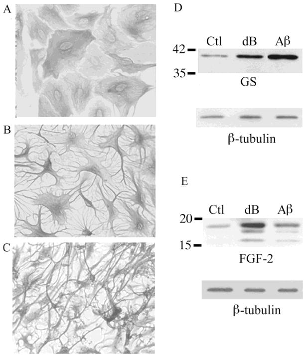
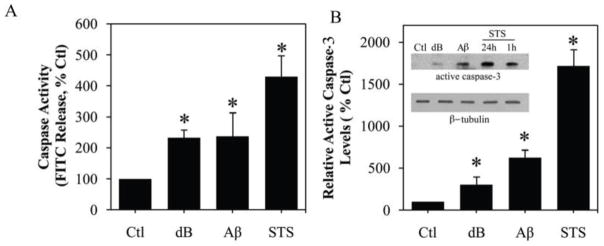
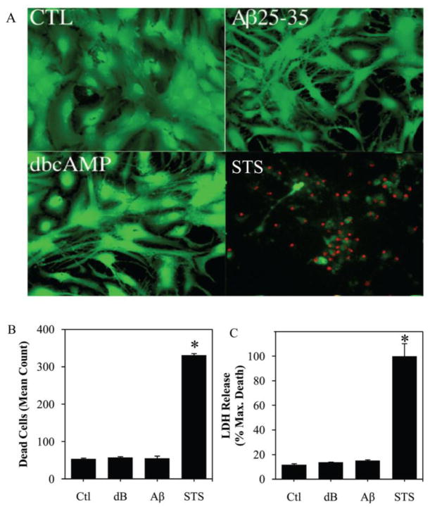
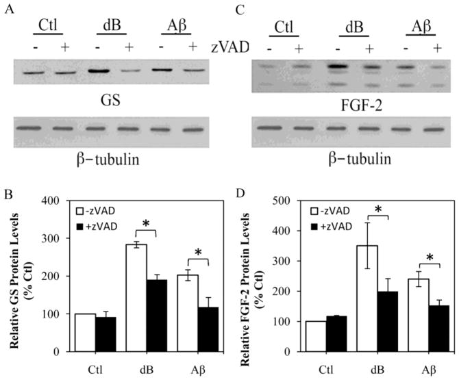
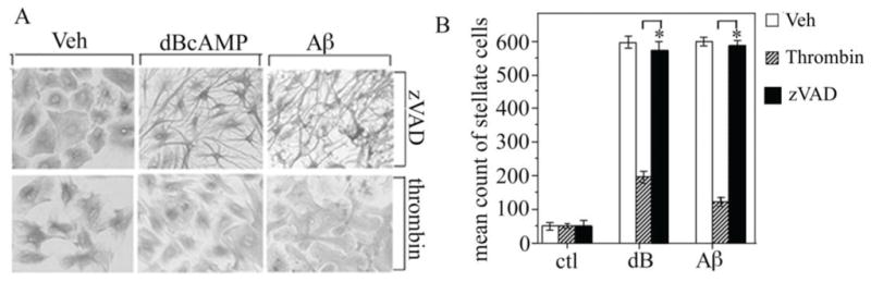
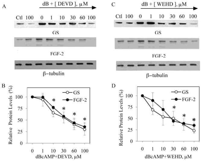
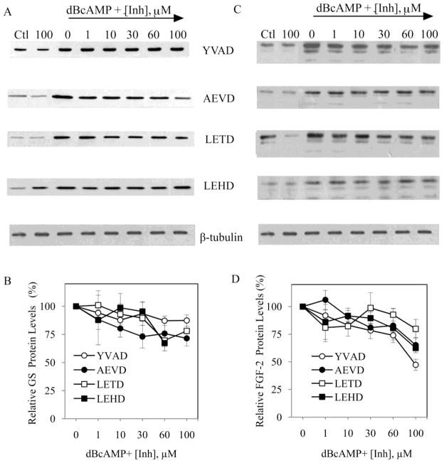
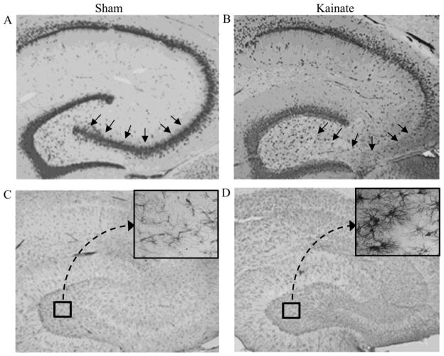
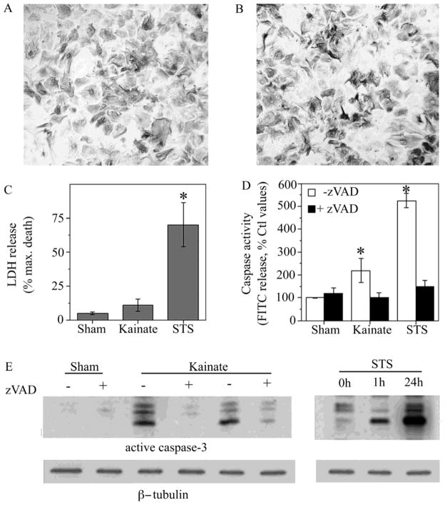
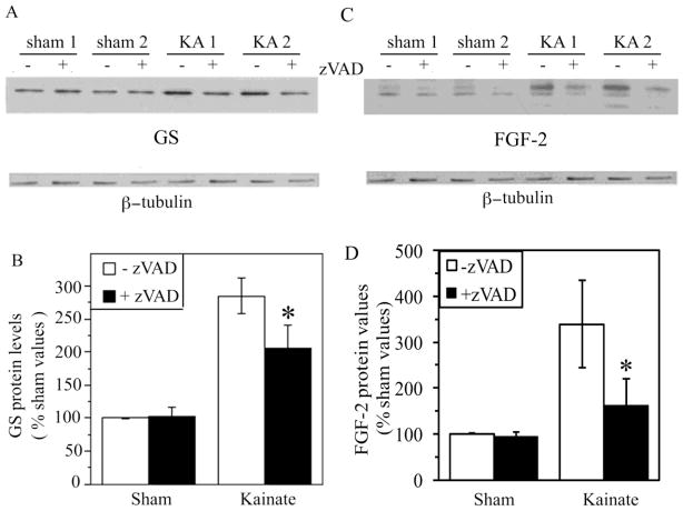
Caspases, a family of cysteine proteases, are widely activated in neurons and glia in the injured brain, a response thought to induce apoptosis. However, caspase activation in astrocytes following injury is not strongly associated with apoptosis. The present study investigates the potential role of caspase activation in astrocytes with another characteristic response to neural injury, astrogliosis. Caspase activity and morphological and biochemical indices of astrogliosis and apoptosis were assessed in (i) cultured neonatal rat astrocytes treated with astrogliosis-inducing stimuli (dibutryl cAMP, β-amyloid peptide), and (ii) cultures of adult rat hippocampal astrocytes generated from control and kainate-lesioned rats. The effects of broad spectrum and specific pharmacological caspase inhibitors were assessed on indicators of astrogliosis, including stellate morphology and expression of glutamine synthetase and fibroblast growth factor-2. Reactive neonatal and adult astrocytes demonstrated an increase in total caspase activity with a corresponding increase in the expression of active caspase-3 in the absence of cell death. Broad spectrum caspase inhibition with zVAD significantly attenuated increases in glutamine synthetase and fibroblast growth factor-2 in the reactive astrocytes. In the reactive neonatal astrocyte cultures, specific inhibition of caspases-3 and -11 also attenuated glutamine synthetase and fibroblast growth factor-2 expression, but did not reverse the morphological reactive phenotype. Astrogliosis is observed in all forms of brain injury and despite extensive study, its molecular triggers remain largely unknown. While previous studies have demonstrated active caspases in astrocytes following acute brain injury, here we present evidence functionally implicating the caspases in astrogliosis.


Figures

Figure 1
Figure 1. Reactivity observed in primary astrocytes treated with dB-cAMP or Aβ
(A) Vehicle-treated control (Ctl) neonatal astrocyte cultures show polygonal morphology. In contrast, stellate morphology is observed in cultures treated with (B) 1 mM dBcAMP (dB) or (C) 25 μM Aβ25–35. Representative western blots demonstrate increased expression of (D) GS and (E) FGF-2 in cultures treated for 48 h with dBcAMP (dB) or Aβ relative to vehicle-treated controls (Ctl).
Figure 2
Figure 2. Increased caspase activity following treatment with reactive stimuli dB-cAMP or Aβ
Treatment of neonatal astrocytes cultures with 1 mM dB-cAMP (dB) or 25 μM Aβ25–35 for 48 h significantly increased (A) total caspase activity and (B) levels of the active caspase-3 fragment as assessed by western blot (inset: representative western blot). Cultures treated for 24 h with the apoptotic stimulus 1μM staurosporine (STS) demonstrated the greatest increase in total caspase activity and levels of the active caspase-3 fragment. Data show mean levels (+SEM) from ≥ 3 independent experiments. * P < 0.01 relative to vehicle-treated control (Ctl).
Figure 3
Figure 3. Absence of cell death following treatment with reactive stimuli db-cAMP or Aβ
(A) Viability of neonatal astrocytes cultures treated with vehicle (CTL), 1 mM dBcAMP or 25 μM Aβ25–35 for 4 d or 1 μM staurosporine (STS) for 24 h was assessed by staining with the vital dyes calcein AM (green fluourescence; indicates live cells), and ethidium homodimer (red fluorescence; indicates dead cells). Levels of cell death in treated astrocytes cultures were quantified by (B) counts of cells with positive staining for ethidium homodimer, and (C) measurement of LDH release into the culture medium. Data show mean levels (+SEM) from ≥ 3 independent experiments. * P < 0.001 relative to vehicle-treated control (Ctl).
Figure 4
Figure 4. Broad-spectrum caspase inhibition attenuates up-regulation of GS and FGF-2 expression in response to reactive stimuli, dB-cAMP and Aβ, in astrocyte cultures
Neonatal astrocyte cultures were treated for 48 h with vehicle (Ctl), 1 mM dBcAMP (dB), or 25 μM Aβ25–35 in the absence (−) or presence (+) or 1 h pretreatment with 50 μM zVAD. Following the experiment, cultures were processed for western blot to assess levels of GS (A) and FGF-2 (C). β-Tubulin was assessed as to ensure equal protein loading. Relative protein levels of GS (B) and FGF-2 (D) were determined by densitometric scanning of western blots from ≥ 3 independent experiments. Data show mean values (+SEM) from each condition expressed as a percentage of the vehicle-treated control group. * P < 0.05 relative to matched (−) zVAD condition.
Figure 5
Figure 5. Astrocyte stellation unaffected by broad spectrum capsase inhibition
Neonatal astrocytes cultures were pretreated for 1 h with vehicle (veh), 50 μM zVAD, or 10 nM thrombin followed by 48 h exposure to 1 mM dBcAMP (dB) or 25 μM Aβ25–35 and then were processed for GFAP immunocytochemistry. (A) Representative bright field images of GFAP immunostaining show that stellate morphology is attenuated by pretreatment with thrombin but not zVAD. (B) Stellation was quantified from immunostained cultures by determining mean counts of morphologically stellate cells (+SEM) from each condition (N = 3). * P < 0.05 relative to matched condition.
Figure 6
Figure 6. Specific inhibition of caspases-3 and -11 attenuates GS and FGF-2 expression in reactive astrocytes
Neonatal astrocytes cultures were pretreated for 1 h with increasing concentration of caspase inhibitors (0, 1, 10, 30, 60, 100 μM) followed by 48 h exposure to 1 mM dBcAMP (dB). For comparison, experiments included conditions with vehicle (Ctl) or 100 μM inhibitor (100) in the absence of dBcAMP. At the conclusion of the experiment, cultures were processed for western blots to assess levels of GS, FGF-2, and β-tubulin (internal control). Representative blots are shown for astrocyte cultures pre-treated with (A) the caspase-3 inhibitor DEVD, and (C) the caspase-11 inhibitor WEHD. Densitometric analysis of blots revealed significant inhibition of both GS and FGF-2 expression in astrocyte cultures treated with (B) 10 – 100 μM DEVD, and (D) 30–100 μM WEHD. Data show mean values (± SEM) relative to dBcAMP-treated cultures in the absence of caspase inhibitor. *P < 0.05 relative to condition lacking inhibitor.
Figure 7
Figure 7. GS and FGF-2 expression in reactive astrocytes unaffected by specific inhibition of caspases-1, -6, -8 and -9
Neonatal astrocytes cultures were pretreated for 1 h with increasing concentration of caspase inhibitors (Inh; 0, 1, 10, 30, 60, 100 μM) followed by 48 h exposure to 1 mM dBcAMP (dB). For comparison, experiments included conditions with vehicle (Ctl) or 100 μM inhibitor (100) in the absence of dBcAMP. At the conclusion of the experiment, cultures were processed for western blots to assess levels of GS, FGF-2, β-tubulin (internal control). Representative blots for (A) GS and (B) FGF-2 are shown for astrocyte cultures pre-treated with specific inhibitors for caspase-1 (YVAD), caspase-6 (AEVD), caspase-8 (LETD) or caspase-9 (LEHD). At non-specific concentrations (>50μM) AEVD and LEHD significantly attenuated both GS and FGF-2 expression, while YVAD attenuated FGF-2 but not GS expression. Data show mean values (± SEM) relative to dBcAMP-treated cultures in the absence of caspase inhibitor.
Figure 8
Figure 8. Kainate lesion results in neuronal loss and astrogliosis
Adult female rats were lesioned bilaterally by intra-hippocampal injection with kainate then 3 weeks later processed for immunohistochemistry. Labeling with the neuron-specific antibody NeuN shows that the hippocampus CA3 pyramidal neuron layer (arrows) is intact in vehicle-lesioned (Sham) animals (A) but largely degenerated in kainate-lesioned (Kainate) rats (B). Astrocyte staining with GFAP antibody shows that, in comparison to sham-lesioned rats (C), lesioned rats display robust labeling throughout hippocampus (D) that is characterized by robust stellation (magnified inset). Images show representative findings from sham-lesioned (N = 4) and kainate-lesioned (N = 5) animals.
Figure 9
Figure 9. Evidence of non-apoptotic caspase activation in an ex vivo model of astrogliosis
Astrocytes cultured from adult sham-lesioned (Sham) and kainate-lesioned (Kainate) rats were characterized for morphology, caspase activity and viability. GFAP immunostaining of cultures revealed similar morphological appearance with little stellation in hippocampal astrocyte cultures derived from (A) sham-lesioned (N = 4) and (B) kainate-lesioned (N = 5) rats. (C) Astrocyte cultures from sham-lesioned animals (Sham) and kainate-lesioned animals (Kainate) did not exhibit significant differences in LDH release. For comparison, cultures from sham-lesioned animals treated for 24 h with 1 μM staurosporine (STS) showed a significant increase in LDH release. (D) Total caspase activity in cultures was assessed in the presence (solid bars) and absence (open bars) of the general caspase inhibitor zVAD (50 μM). Caspase activity was significantly elevated both in cultures from kainate-lesioned animals and in cultures of sham-lesioned animals treated for 24 h with 1 μM staurosporine. Data show mean values (+SEM) from 4–5 independent culture preparations. * P < 0.05 relative to Sham condition. (E) Representative western blots show relative levels active caspase-3 expression from sham- and kainate-lesioned astrocyte cultures in the presence (+) and absence (−) of 50 μM zVAD treatment and following treatment with 1μM staurosporine (STS) for 1 h or 24 h. Bots were stripped and reprobed β-tubulin as an internal control for equal protein.
Figure 10
Figure 10. Caspase inhibition attenuates up-regulation of GS and FGF-2 expression in an ex vivo model of astrogliosis
Astrocyte cultures from sham- and kainate-lesioned rats were treated for 48 h with vehicle (−) or 50 μM general caspase inhibitor zVAD (+) and then processed for western blot. Representative western blots show increased (A) GS and (C) FGF-2 expression in cultures from kainate-lesioned (KA 1, KA 2) rats compared to sham-lesioned controls (Sham 1, Sham 2). Densitometric analysis of western blots revealed zVAD treatment (+zVAD; solid bars) significantly decreased (B) GS and (D) FGF-2 expression in astrocyte cultures from Kainate-lesioned rats (Sham, N = 4; Kainate, N = 5). * P < 0.05 relative to matched condition.

References

    1. Abe K, Kato M, Saito H. Human amylin mimicks amyloid [beta] protein-induced reactive gliosis and inhibition of cellular redox activity in cultured astrocytes. Brain Res. 1997;762:285–288. - PubMed
    1. Abe K, Saito H. Na+ and K+ dependence of morphological changes of cultured rat cortical astrocytes. Pharmacol Toxicol. 2001;88:319–24. - PubMed
    1. Abe K, Saito H. The p44/42 mitogen-activated protein kinase cascade is involved in the induction and maintenance of astrocyte stellation mediated by protein kinase C. Neurosci Res. 2000;36:251–7. - PubMed
    1. Acarin L, Villapol S, Faiz M, Rohn TT, Castellano B, González B. Caspase-3 activation in astrocytes following postnatal excitotoxic damage correlates with cytoskeletal remodeling but not with cell death or proliferation. Glia. 2007;55:954–965. - PubMed
    1. Araujo DM, Cotman CW. [beta]-Amyloid stimulates glial cells in vitro to produce growth factors that accumulate in senile plaques in Alzheimer’s disease. Brain Res. 1992;569:141–145. - PubMed

Publication types

MeSH terms