. 2012 May;91(5):454-9.
doi: 10.1177/0022034512443068. Epub 2012 Mar 20.

Dentin-cement interfacial interaction: calcium silicates and polyalkenoates

Affiliations

Dentin-cement interfacial interaction: calcium silicates and polyalkenoates

A R Atmeh et al. J Dent Res. 2012 May.

Abstract

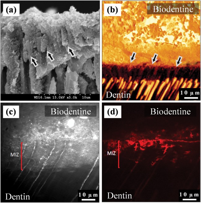
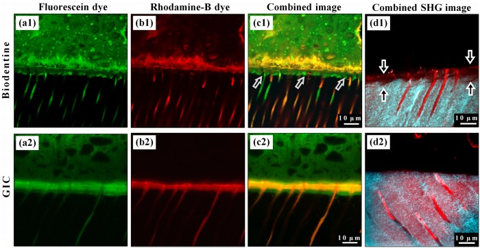
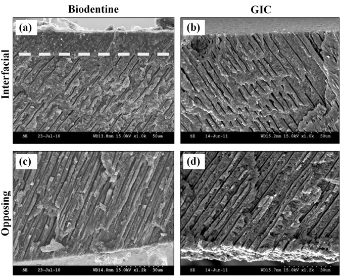
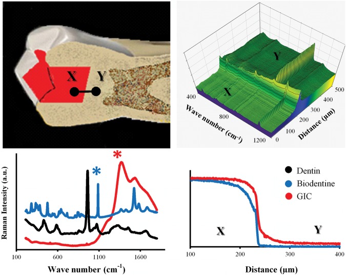
The interfacial properties of a new calcium-silicate-based coronal restorative material (Biodentine™) and a glass-ionomer cement (GIC) with dentin have been studied by confocal laser scanning microscopy (CLSM), scanning electron microscopy (SEM), micro-Raman spectroscopy, and two-photon auto-fluorescence and second-harmonic-generation (SHG) imaging. Results indicate the formation of tag-like structures alongside an interfacial layer called the "mineral infiltration zone", where the alkaline caustic effect of the calcium silicate cement's hydration products degrades the collagenous component of the interfacial dentin. This degradation leads to the formation of a porous structure which facilitates the permeation of high concentrations of Ca(2+), OH(-), and CO(3) (2-) ions, leading to increased mineralization in this region. Comparison of the dentin-restorative interfaces shows that there is a dentin-mineral infiltration with the Biodentine, whereas polyacrylic and tartaric acids and their salts characterize the penetration of the GIC. A new type of interfacial interaction, "the mineral infiltration zone", is suggested for these calcium-silicate-based cements.


Conflict of interest statement

The author(s) declare no potential conflicts of interest with respect to the authorship and/or publication of this article.

Figures

Figure 1.
Figure 1.
Interfacial characteristics. (a) SEM micrograph of fractured dentin beneath a Biodentine restoration. Tag-like structures were detected forming within the dentinal tubules (arrows). (b) Fluorescence-mode CLSM image showing the cement tags, which appear on the interfacial surface of the fluorescein-labeled Biodentine (above) after it was pulled away from dentin due to desiccation. 63x/1.4NA OI. (c) Reflection-mode TSM image for the dentin/Biodentine interface. The mineral infiltration zone (MIZ) appears as a band of highly reflective dentin beneath the interface, indicating a change in dentin’s mineral content within this zone. The fluorescence-mode image of the same area (d) shows the distribution of Rhodamine-B dye, which permeated from the pulp chamber into the interface and cement.
Figure 2.
Figure 2.
Representative fluorescence-mode CLSM images for the interfaces of Biodentine and GIC cements (upper side of each image) with the dentin (lower side). (a1) Distribution of the fluorescein dye labeling the Biodentine cement shows a band of richly dye-infiltrated dentin just beneath the interface. This band cannot be seen with Rhodamine-B micro-permeability (b1), where the dye permeated through this band and diffused into the cement above the interface without any mixing with the fluorescein. This band is believed to be formed as a result of the strong alkaline effect of calcium hydroxide leaching out of the cement, and it corresponds to the MIZ. (c1) Dye-deficient areas can be seen within this zone (arrows), which represent the un-affected peritubular dentin. (a2) Fluorescein distribution in GIC samples also shows a richly dye-infiltrated dentin band, but this band was formed due to the acidic effect of GIC, which affected both intertubular and peritubular dentin. (b2) Rhodamine-B permeating from the pulp has also infiltrated this band, which diffused laterally through the affected tubular walls and mixed with the fluorescein as shown in (c2). (d1) Second-harmonic-generation (SHG) signal, originating from the intertubular collagen (cyan), is weak and almost absent in the interfacial dentin beneath the Biodentine (arrows) when superimposed over the dentin’s autofluorescence signal (red), which reflects its actual margin. Notice the preserved tubular structure of dentin in this zone, unlike the images for GIC samples (d2), where the SHG signal has the same distribution as the dentin’s autofluorescence signal.
Figure 3.
Figure 3.
SEM micrographs of the fractured dentin discs which were exposed to Biodentine (a) or GIC (b). A band of structurally altered dentin extends along the interface, as can be seen by the obliterated dentin tubules above the dotted line in (a), when compared with dentin of the opposite surface of the disc, where no cement was applied (c). (b) No structural changes can be detected in the dentin beneath GIC when compared with dentin of the opposite surface (d).
Figure 4.
Figure 4.
(upper left) Raman area maps were obtained for the dentin-cement interface in GIC and Biodentine filled teeth by StreamLine™ scanning in the direction from points X to Y. Raman spectral maps were analyzed with curve-fitting software which averaged each area map into a single line, where each point on that line was represented by an averaged Raman spectrum (upper right). From the cements’ Raman spectra (lower left), representative characteristic peaks [1082 cm-1 and 1262 cm-1 (marked with asterisks)] were designated for Biodentine and GI cements, respectively. The average penetration depth was derived from the profiles of normalized Raman intensity of each peak, after they were generated from the curve-fitting software (lower right).

References

    1. Andersen M, Lund A, Andreasen JO, Andreasen FM. (1992). In vitro solubility of human pulp tissue in calcium hydroxide and sodium hypochlorite. Dent Traumatol 8:104-108 - PubMed
    1. Andreasen JO, Farik B, Munksgaard EC. (2002). Long-term calcium hydroxide as a root canal dressing may increase risk of root fracture. Dent Traumatol 18:134-137 - PubMed
    1. Bowes JH. (1950). The swelling of collagen in alkaline solutions; swelling in solutions of bivalent bases. Biochem J 46:530-532 - PMC - PubMed
    1. Camilleri J, Montesin FE, Papaioannou S, McDonald F, Pitt Ford TR. (2004). Biocompatibility of two commercial forms of mineral trioxide aggregate. Int Endod J 37:699-704 - PubMed
    1. Camilleri J, Cutajar A, Mallia B. (2011). Hydration characteristics of zirconium oxide replaced Portland cement for use as a root-end filling material. Dent Mater 27:845-854 - PubMed

Publication types

LinkOut - more resources