. 2012 Mar 21;485(7398):327-32.
doi: 10.1038/nature10939.

Structure of the human κ-opioid receptor in complex with JDTic

Affiliations

Structure of the human κ-opioid receptor in complex with JDTic

Huixian Wu et al. Nature. .

Abstract

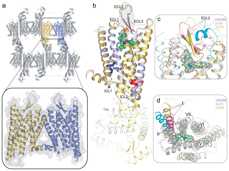
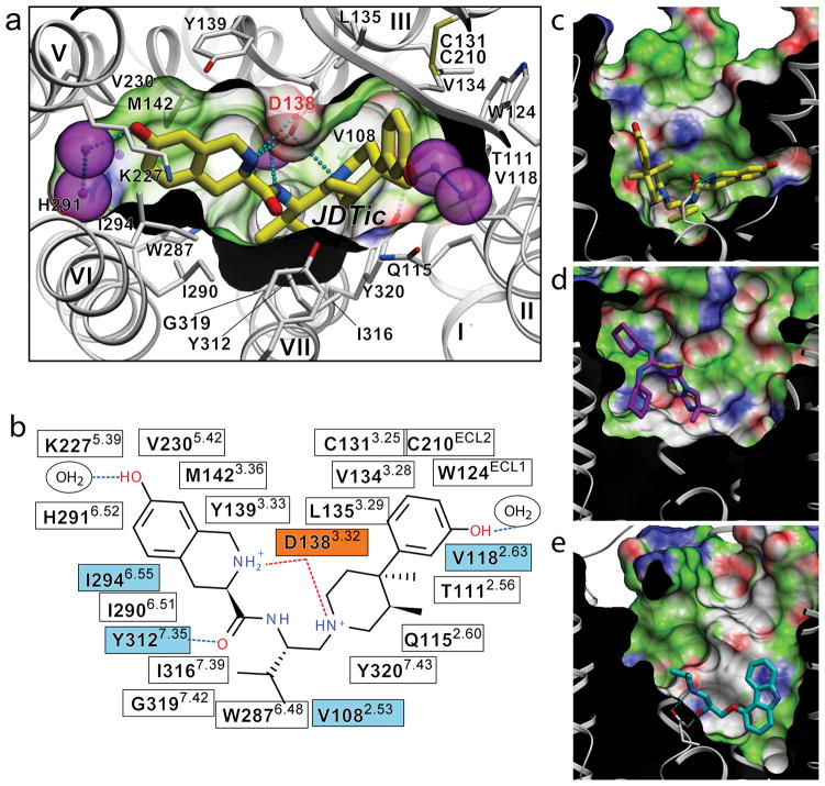
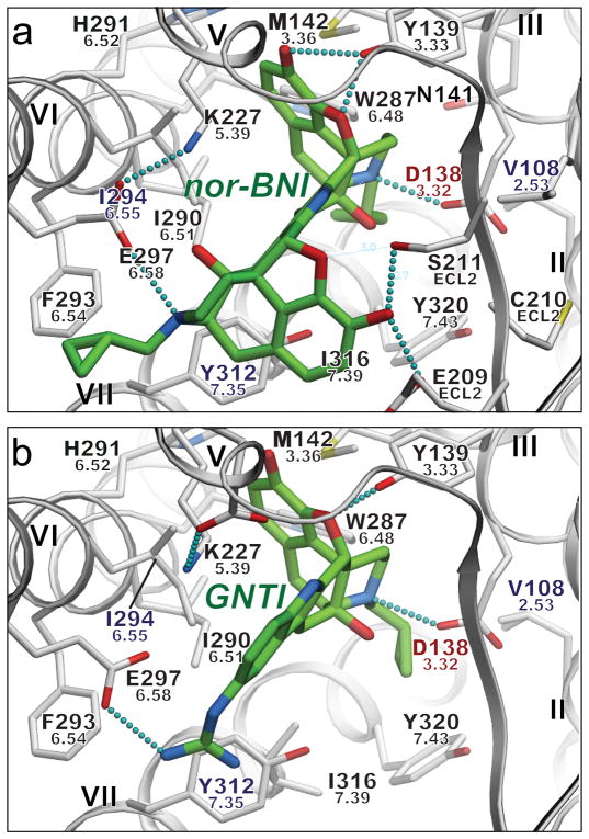
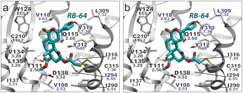
Opioid receptors mediate the actions of endogenous and exogenous opioids on many physiological processes, including the regulation of pain, respiratory drive, mood, and--in the case of κ-opioid receptor (κ-OR)--dysphoria and psychotomimesis. Here we report the crystal structure of the human κ-OR in complex with the selective antagonist JDTic, arranged in parallel dimers, at 2.9 Å resolution. The structure reveals important features of the ligand-binding pocket that contribute to the high affinity and subtype selectivity of JDTic for the human κ-OR. Modelling of other important κ-OR-selective ligands, including the morphinan-derived antagonists norbinaltorphimine and 5'-guanidinonaltrindole, and the diterpene agonist salvinorin A analogue RB-64, reveals both common and distinct features for binding these diverse chemotypes. Analysis of site-directed mutagenesis and ligand structure-activity relationships confirms the interactions observed in the crystal structure, thereby providing a molecular explanation for κ-OR subtype selectivity, and essential insights for the design of compounds with new pharmacological properties targeting the human κ-OR.


Figures

Figure 1
Figure 1. Crystal packing and overview of the hKOR structure in complex with JDTic, and comparison with the inactive CXCR4 and β2AR structures
(a) hKOR-T4L crystal packing. The parallel-dimer in one ASU is highlighted by insert. (b) Overall architecture of hKOR-T4L in complex with JDTic. A molecule (yellow) and B molecule (blue) in one ASU are aligned through the receptor part. The DRY and NPxxY motifs are highlighted in red and blue, respectively. JDTic is shown in a green sphere representation and the disulfide bonds are colored orange. (c) Side and (d) extracellular views of a structural alignment of hKOR (yellow); CXCR4 (PDB ID: 3ODU; magenta) and β2AR (PDB ID: 2RH1; cyan). The graphics were created by PyMOL.
Figure 2
Figure 2. Binding of the high affinity selective antagonist JDTic in the hKOR crystal structure
(a) Conformation of the binding pocket with JDTic shown by sticks with yellow carbons. The protein is displayed in cartoon representation looking down from the extracellular side, with the 22 contact residues within 4.5 Å from the ligand shown by white sticks. The pocket surface is shown as a semitransparent surface colored according to binding properties (green: hydrophobic, blue: H-bond donor, red: H-bond acceptor). Salt bridges and hydrogen bonds are shown as dotted lines. Structured water molecules are shown as large magenta spheres. (b) Diagram of ligand interactions in the binding pocket side chains at 4.5 Å cutoff. Salt bridges are shown in red and direct hydrogen bonds in blue dashed lines. Ballesteros-Weinstein numbering is shown as superscript. Residues that vary among MOR, DOR and KOR subtypes are highlighted in cyan, and residue Asp1383.32 inferred in hKOR ligand binding by mutagenesis data, is highlighted orange. Side views of the sliced binding pocket in (c) hKOR-JDTic, (d) CXCR4-IT1t, and (e) β2AR-carazolol complexes. The pocket surfaces are colored as in panel A, the protein interior is black and the extracellular space is white. Ligands are shown as capped sticks with carbons colored yellow (JDTic), magenta (IT1t) and cyan (carazolol). Asp3.32 side chains in hKOR-JDTic and β2AR-carazolol complexes are shown by thin sticks with grey carbons. The graphics were prepared using ICM molecular modeling package (Molsoft LLC).
Figure 3
Figure 3. Putative interaction modes of morphine-based high affinity hKOR selective antagonists nor-BNI (a) and GNTI (b)
Ligands are depicted as capped sticks with green carbons, and contact side chains of the receptor within 4 Å from the ligand are shown with grey carbons. Key hydrogen bonds and salt bridges are indicated with small cyan spheres and residues unique to KOR are labeled in blue. Residue Asp1383.32, which also shows critical impact on GNTI and nor-BNI binding in mutagenesis studies, is highlighted red. Ballesteros-Weinstein residue numbers are shown under the hKOR residue numbers. The graphics were prepared using ICM molecular modeling package (Molsoft LLC).
Figure 4
Figure 4. Model of covalently-bound RB-64
Putative binding mode of (a) the RB-64 +463 amu and (b) the RB-64 +431 amu adduct. Residues within 4 Å of the ligand are displayed. Rendering: ligand, capped sticks/cyan carbons; hKOR side chains, capped sticks; hydrogen bonds, small green spheres; hKOR-unique residues labeled in blue. Ballesteros-Weinstein residue numbers are shown under the hKOR residue numbers. The graphics were prepared using ICM molecular modeling package (Molsoft LLC).

Comment in

References

    1. Fredriksson R, Lagerstrom MC, Lundin LG, Schioth HB. The G-protein-coupled receptors in the human genome form five main families. Phylogenetic analysis, paralogon groups, and fingerprints. Mol Pharmacol. 2003;63:1256–1272. - PubMed
    1. Waldhoer M, Bartlett SE, Whistler JL. Opioid receptors. Annu Rev Biochem. 2004;73:953–990. - PubMed
    1. Cherezov V, et al. High-resolution crystal structure of an engineered human beta2-adrenergic G protein-coupled receptor. Science. 2007;318:1258–1265. - PMC - PubMed
    1. Jaakola VP, et al. The 2.6 angstrom crystal structure of a human A2A adenosine receptor bound to an antagonist. Science. 2008;322:1211–1217. - PMC - PubMed
    1. Chien EY, et al. Structure of the human dopamine D3 receptor in complex with a D2/D3 selective antagonist. Science. 2010;330:1091–1095. - PMC - PubMed

Publication types

MeSH terms

Substances

Associated data