

Biceps femoris muscle transposition for treatment of cranial cruciate ligament rupture in small breed dogs
- PMID: 22437541
- PMCID: PMC3317463
- DOI: 10.4142/jvs.2012.13.1.93
Biceps femoris muscle transposition for treatment of cranial cruciate ligament rupture in small breed dogs
Abstract
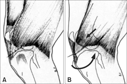
The purpose of this study was to evaluate a new extracapsular surgical technique for the treatment of cranial cruciate ligament rupture in small breed dogs. Nine small breed dogs (seven females and two males) weighing ≤ 15 kg were treated with biceps femoris muscle transposition (BFT). The duration of the BFT procedure was 20 min. Each patient underwent a standard clinical protocol and a questionnaire for the owners. Follow-up (at 1, 3, and 12 months postoperative) confirmed significant improvement in all patients, especially at 1 month postoperatively (p < 0.01) and again after complete stifle joint assessment at 3 months postoperatively. After 12 months, only two patients showed a slight increase in osteoarthritis. According to our results, BFT is a simple extracapsular surgical technique that can be used for the treatment of cranial cruciate ligament rupture in small breed dogs.
Figures


References
-
- Aragon CL, Budsberg SC. Applications of evidence-based medicine: cranial cruciate ligament injury repair in the dog. Vet Surg. 2005;34:93–98. - PubMed
-
- Arnoczky SP, Tarvin GB, Marshall JL, Saltzman B. The over the top procedure: a technique for anterior cruciate ligament substitution in the dog. J Am Anim Hosp Assoc. 1979;15:283–290.
-
- Arthurs GI, Langley-Hobbs SJ. Patellar luxation as a complication of surgical intervention for the management of cranial cruciate ligament rupture in dogs. A retrospective study of 32 cases. Vet Comp Orthop Traumatol. 2007;20:204–210. - PubMed
-
- Brown DC. Outcomes based medicine in veterinary surgery: getting hard measures of subjective outcomes. Vet Surg. 2007;36:289–292. - PubMed
-
- Cook JL, Luther JK, Beetem J, Karnes J, Cook CR. Clinical comparison of a novel extracapsular stabilization procedure and tibial plateau leveling osteotomy for treatment of cranial cruciate ligament deficiency in dogs. Vet Surg. 2010;39:315–323. - PubMed
MeSH terms
LinkOut - more resources
Full Text Sources
