. 2013 Aug;34(8):1768-82.
doi: 10.1002/hbm.22035. Epub 2012 Mar 22.

Brain activity during sympathetic response in anticipation and experience of pain

Affiliations

Brain activity during sympathetic response in anticipation and experience of pain

Frank Seifert et al. Hum Brain Mapp. 2013 Aug.

Abstract

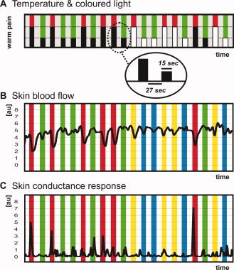
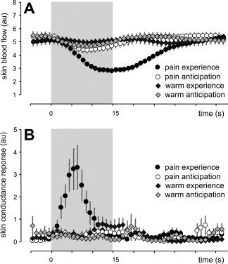
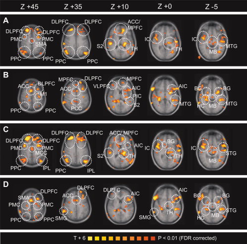
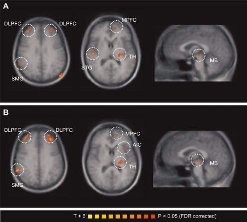
Pain is a multidimensional phenomenon with sensory, affective, and autonomic components. Here, we used parametric functional magnetic resonance imaging (fMRI) to correlate regional brain activity with autonomic responses to (i) painful stimuli and to (ii) anticipation of pain. The autonomic parameters used for correlation were (i) skin blood flow (SBF) and (ii) skin conductance response (SCR). During (i) experience of pain and (ii) anticipation of pain, activity in the insular cortex, anterior cingulate cortex (ACC), prefrontal cortex (PFC), posterior parietal cortex (PPC), secondary somatosensory cortex (S2), thalamus, and midbrain correlated with sympathetic outflow. A conjunction analysis revealed a common central sympathetic network for (i) pain experience and (ii) pain anticipation with similar correlations between brain activity and sympathetic parameters in the anterior insula, prefrontal cortex, thalamus, midbrain, and temporoparietal junction. Therefore, we here describe shared central neural networks involved in the central autonomic processing of the experience and anticipation of pain.

Keywords: autonomic nervous system; fMRI; neuropathic pain; pain; somatosensory system.


Figures

Figure 1
Figure 1
Experimental procedure. A: Schematic diagram and time‐line of the applied stimuli. Visually displayed colored lights signaled the heat pain (red light) and the innocuous warmth (green light) stimuli. After conditioning, only the light was displayed, but no thermal stimulus was applied (indicated by white boxes). B: The mean SBF during the experiment is shown: heat pain experience (red bars), warmth experience (green bars), heat pain anticipation (yellow bars), and warmth anticipation (blue bars). C: The mean skin conduction response during the experiment is shown: heat pain experience (red bars), warmth experience (green bars), heat pain anticipation (yellow bars), and warmth anticipation (blue bars). [Color figure can be viewed in the online issue, which is available at wileyonlinelibrary.com.]
Figure 2
Figure 2
A: Skin blood flow during all tested conditions. B: Skin resistance during all tested conditions. Means ± SEM.
Figure 3
Figure 3
Parametrical fMRI analysis. The T‐statistic contrast map shows areas with activity covarying with autonomic response: (A) SBF and heat pain (heat pain–warmth), (B) SBF and anticipation of heat pain (pain anticipation–anticipation of warmth), (C) SCR and heat pain (heat pain–warmth), (D) SCR and anticipation of pain (pain anticipation–anticipation of warmth). The group statistic contrast maps are registered onto the averaged Talairach‐transformed brains, thresholded at P < 0.01 (FDR corrected). The Talairach‐coordinates and cluster sizes are depicted in Table I. Left hemisphere—ipsilateral; right hemisphere—contralateral. ACC, anterior cingulate cortex; MPFC, medial prefrontal cortex; DLPFC, dorsolateral prefrontal cortex; VLPFC, ventrolateral prefrontal cortex; IC, insular cortex; AIC, anterior insular cortex; PIC, posterior insular cortex; S2, secondary somatosensory cortex; PPC, posterior parietal cortex; M1, primary motor cortex; PMC; premotor cortex; MTG, middle temporal gyrus; STG, superior temporal gyrus; IPL, inferior parietal lobule; HC, hippocampus; BG, basal ganglia; TH, thalamus; MB, midbrain. [Color figure can be viewed in the online issue, which is available at wileyonlinelibrary.com.]
Figure 4
Figure 4
Conjunction analysis of the parametrical fMRI data. The T‐statistic conjunction map shows areas in which activity covaries with autonomic response similarly during pain experience and pain anticipation: (A) skin blood flow and (B) sympathetic skin response. The group statistic conjunction maps are registered onto the averaged Talairach‐transformed brains, thresholded at P < 0.05 (FDR corrected). The Talairach‐coordinates and cluster sizes are depicted in Table II. Left hemisphere—ipsilateral; right hemisphere—contralateral. MPFC, medial prefrontal cortex; DLPFC, dorsolateral prefrontal cortex; VLPFC, ventrolateral prefrontal cortex; AIC, anterior insular cortex; MTG, middle temporal gyrus; STG, superior temporal gyrus; TH, thalamus; MB, midbrain. [Color figure can be viewed in the online issue, which is available at wileyonlinelibrary.com.]

References

    1. Apkarian AV, Bushnell MC, Treede RD, Zubieta JK ( 2005): Human brain mechanisms of pain perception and regulation in health and disease. Eur J Pain 9: 463–484. - PubMed
    1. Bantick SJ, Wise RG, Ploghaus A, Clare S, Smith SM, Tracey I ( 2002): Imaging how attention modulates pain in humans using functional MRI. Brain 125( Part 2): 310–319. - PubMed
    1. Bechara A, Tranel D, Damasio H ( 2000): Characterization of the decision‐making deficit of patients with ventromedial prefrontal cortex lesions. Brain 123( Part 11): 2189–2202. - PubMed
    1. Benarroch EE ( 2006): Pain‐autonomic interactions. Neurol Sci 27( Suppl 2): S130–S133. - PubMed
    1. Bingel U, Lorenz J, Schoell E, Weiller C, Buchel C ( 2006): Mechanisms of placebo analgesia: rACC recruitment of a subcortical antinociceptive network. Pain 120: 8–15. - PubMed

Publication types