. 2012 Jun;86(11):6258-67.
doi: 10.1128/JVI.00189-12. Epub 2012 Mar 21.

Molecular characterization of feline infectious peritonitis virus strain DF-2 and studies of the role of ORF3abc in viral cell tropism

Affiliations

Molecular characterization of feline infectious peritonitis virus strain DF-2 and studies of the role of ORF3abc in viral cell tropism

Ádám Bálint et al. J Virol. 2012 Jun.

Abstract

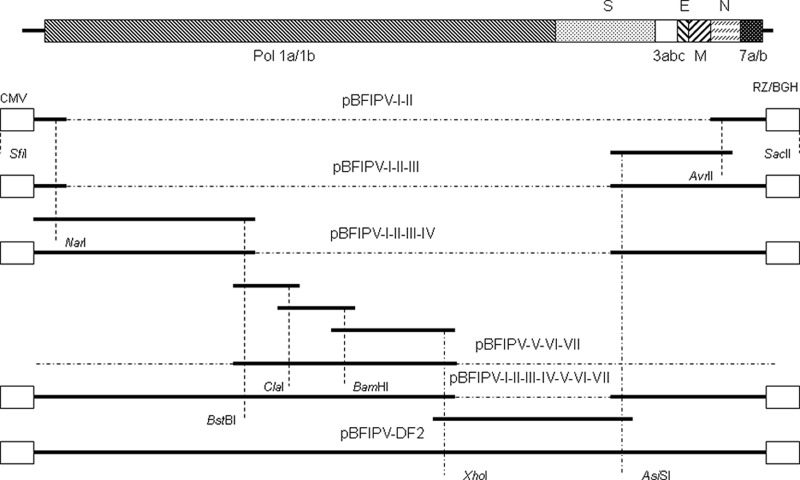
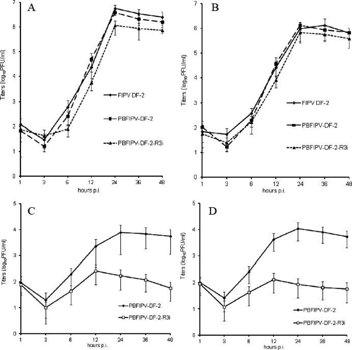
The full-length genome of the highly lethal feline infectious peritonitis virus (FIPV) strain DF-2 was sequenced and cloned into a bacterial artificial chromosome (BAC) to study the role of ORF3abc in the FIPV-feline enteric coronavirus (FECV) transition. The reverse genetic system allowed the replacement of the truncated ORF3abc of the original FIPV DF-2 genome with the intact ORF3abc of the canine coronavirus (CCoV) reference strain Elmo/02. The in vitro replication kinetics of these two viruses was studied in CrFK and FCWF-4 cell lines, as well as in feline peripheral blood monocytes. Both viruses showed similar replication kinetics in established cell lines. However, the strain with a full-length ORF3 showed markedly lower replication of more than 2 log(10) titers in feline peripheral blood monocytes. Our results suggest that the truncated ORF3abc plays an important role in the efficient macrophage/monocyte tropism of type II FIPV.


Figures

Fig 1
Fig 1
Cloning strategy for the FIPV DF-2 genome. Viral genes are represented by shaded boxes on the genome. Open boxes represent the 5′ and 3′ accessory sequences of the vector. Genome parts are represented with continuous boldface lines.
Fig 2
Fig 2
Recombination map of FIPV DF-2. (A) SimPlot analysis of the FIPV DF-2 genome. FIPV DF-2 was compared to the reference strains FCoV UU8 and CCoV NTU336. Percent identity is represented on the y axis, and nucleotide positions are shown on the x axis. The sliding window was selected as 200 nt, with a step size of 20 nt. (B) Sequence alignment flanking the putative recombination sites of FIPV DF-2. FCoV UU8 and FCoV Black are type I FCoVs, and FIPV DF-2 and FIPV WSU 79-1146 are type II FCoVs. NTU 336 is a CCoV strain. Only sequences flanking the putative donor (DS) and acceptor (AS) recombination sites of FIPV DF-2 are shown. Nucleotides different from the consensus type I sequence are shaded by different tones of gray. Regions where the recombination most probably could have taken place are shaded black.
Fig 3
Fig 3
Schematic representation of region 3 of the CCoV Elmo/02 (A) and FIPV DF-2 (B) strains. F1 to F3 represent the three reading frames of the viral genomes. Numbering starts at the first nucleotide of the regions' first core sequence (CUAAAC). The ORFs of interest are symbolized by shaded boxes and are labeled by the number of nucleotides at the beginning and the end. Dashed lines symbolize the first start codon in the ORFs. The dashed connector line represents the deleted part in the DF-2 genome. Numbers in brackets correspond to the numbering of the appropriate nucleotides in region 3 of CCoV Elmo/02.
Fig 4
Fig 4
Alignment and topology prediction of the most divergent part of the nsp6 proteins of the DF-2 and DF-2 vaccine strains. Topology predictions were executed by the TMpred and HMMTOP programs on the full nsp6 proteins; however, only the sites of interest are shown (I, inside loop; o, outside loop; H, transmembrane helix). Numbers indicate the positions of the amino acids in the 1a polyproteins.
Fig 5
Fig 5
Replication dynamics of the different FCoVs. Growth kinetics of the PBFIPV-DF-2 and PBFIPV-DF-2-R3i recombinant viruses and the parent FIPV DF-2 strain after infection of FCWF-4 (A) and CrFK (B) cells (MOI of 0.1). The titers are given as the means from triplicate experiments (log10 TCID50/ml); error bars represent standard deviations. Also shown are the growth kinetics of intracellular (C) and extracellular (D) recombinant viruses PBFIPV-DF-2 and PBFIPV-DF-2-R3i after the infection of feline monocytes originating from five different cats (MOI = 5).
Fig 6
Fig 6
RT-PCR detection of sg mRNA transcription of ORF3abc of PBFIPV-DF-2 and PBFIPV-DF-2-R3i 24 h after infection of CrFK cells (MOI of 0.1). The DF2F forward primer was designed to hybridize to the leader sequence of the FIPV DF-2 genome, while the ORF3CR reverse primer annealed to a sequence stretch of ORF3c present in both viruses. Lane 1, amplification of the ORF3abc sg RNA of PBFIPV-DF-2 resulting in a 804-bp-long amplicon corresponding to the truncated ORF3abc sg mRNA. Lane 2, amplification of the ORF3abc sg RNAs of PBFIPV-DF-2-R3i resulting in three amplicons: a 1,142-bp-long product corresponding to the intact ORF3abc sg mRNA, a 950-bp amplicon corresponding to the ORF3bc sg mRNA, and a 758-bp fragment corresponding to ORF3c sg mRNA. Lane 3, GeneRuler 1-kb Plus DNA ladder (Fermentas).
Fig 7
Fig 7
Quantification of genomic and replicative RNA. Copy numbers of FIPV DF2 and recombinant viruses PBFIPV-DF-2 and PBFIPV-DF-2-R3i at 24 h p.i. in FCWF-4 cells, CrFK cells (MOI = 0.1), and feline monocytes originating from five different cats (MOI of 5) with genomic (A) and subgenomic (B) TaqMan assays.

References

    1. Addie D, Jarrett O. 1992. A study of naturally occurring feline coronavirus infections in kittens. Vet. Rec. 130:133–137 - PubMed
    1. Addie D, et al. 2009. Feline infectious peritonitis. ABCD guidelines on prevention and management. J. Feline Med. Surg. 11:594–604 - PMC - PubMed
    1. Almazan F, et al. 2000. Engineering the largest RNA virus genome as an infectious bacterial artificial chromosome. Proc. Natl. Acad. Sci. U. S. A. 97:5516–5521 - PMC - PubMed
    1. Almazan F, Galan C, Enjuanes L. 2004. The nucleoprotein is required for efficient coronavirus genome replication. J. Virol. 78:12683–12688 - PMC - PubMed
    1. Almazan F, et al. 2006. Construction of a severe acute respiratory syndrome coronavirus infectious cDNA clone and a replicon to study coronavirus RNA synthesis. J. Virol. 80:10900–10906 - PMC - PubMed

Publication types

Associated data

LinkOut - more resources