. 2012 May;23(10):1812-25.
doi: 10.1091/mbc.E11-12-1006. Epub 2012 Mar 21.

Reggies/flotillins regulate E-cadherin-mediated cell contact formation by affecting EGFR trafficking

Affiliations

Reggies/flotillins regulate E-cadherin-mediated cell contact formation by affecting EGFR trafficking

Gonzalo P Solis et al. Mol Biol Cell. 2012 May.

Abstract

The reggie/flotillin proteins are implicated in membrane trafficking and, together with the cellular prion protein (PrP), in the recruitment of E-cadherin to cell contact sites. Here, we demonstrate that reggies, as well as PrP down-regulation, in epithelial A431 cells cause overlapping processes and abnormal formation of adherens junctions (AJs). This defect in cell adhesion results from reggie effects on Src tyrosine kinases and epidermal growth factor receptor (EGFR): loss of reggies reduces Src activation and EGFR phosphorylation at residues targeted by Src and c-cbl and leads to increased surface exposure of EGFR by blocking its internalization. The prolonged EGFR signaling at the plasma membrane enhances cell motility and macropinocytosis, by which junction-associated E-cadherin is internalized and recycled back to AJs. Accordingly, blockage of EGFR signaling or macropinocytosis in reggie-deficient cells restores normal AJ formation. Thus, by promoting EGFR internalization, reggies restrict the EGFR signaling involved in E-cadherin macropinocytosis and recycling and regulate AJ formation and dynamics and thereby cell adhesion.


Figures

FIGURE 1:
FIGURE 1:
Reggies regulate cell contact inhibition and intercellular adhesion in A431 cells. Immunostaining of endogenous PrP (A) and reggie-1 (R1, B) showed that both proteins colocalized with E-cadherin (E-cad) at cell contact sites in A431 cells. Scale bars, 10 μm. (C) Down-regulation of reggie-1 (shR1) or PrP (shPrP) induced a significant increase in overlapping processes (yellow arrowheads) as revealed by E-cad immunostaining. Scale bars, 10 μm. (D) Quantification of overlapping areas (n = 3, ***p < 0.001, one-way ANOVA, mean ± SEM). (E) Intercellular adhesion was analyzed using the dispase-based dissociation assay. Whereas a low degree of fragmentation of the cell carpets was observed in control shLuc cells (left), increased levels of fragmentation were apparent in shR1 (middle) and shPrP (right) cells. (F) Quantification of the dispase assay (n = 4, **p < 0.01, ***p < 0.001, one-way ANOVA, mean ± SEM).
FIGURE 2:
FIGURE 2:
Disruption of AJ formation and dynamics after reggie-1 and PrP down-regulation. (A) Immunostainings of the detergent-resistant pools of E-cadherin (E-cad) and β-catenin (β-cat) showed the streaks typical of AJs at contact sites in control A431 cells (shLuc, first row) and revealed disorganized AJs in reggie-1 (shR1, second row) and PrP (shPrP, third row) knockdown cells (enlargement of boxed areas on the right). Scale bars, 10 μm. (B) E-cadherin–EGFP (E-cad-EGFP)–expressing cells were transfected with control siRNA (siGL2), siRNA against reggie-1 (siR1), or siRNA against PrP (siPrP) and AJ movements recorded for 20 min. In contrast to control cells (left), AJs were not well defined, and their basal-to-apical movements were significantly reduced in siR1 (middle) and siPrP (right) cells. Trajectories of individual AJs from basal (b) to apical (a) regions of overlapping cell contacts (outlined by black lines) are shown in color (bottom). Scale bars, 5 μm. (C, D) Quantifications showed a significant reduction of velocity (C) and number (D) of AJs in cells treated as in B (n = 5, *p < 0.05, ***p < 0.001, one-way ANOVA, mean ± SEM).
FIGURE 3:
FIGURE 3:
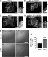
EGFR blocker rescued AJ formation defects of shR1 cells. (A–D) E-cadherin–EGFP (E-cad-EGFP) expression in shRNA stably transfected A431 cells revealed the reduction in AJ formation (boxed area enlarged in inserts) and increased cell motility (white lines and kymographs on the right) in shR1 cells (C) compared with control shLuc cells (A). Stimulation of shLuc cells with 10 ng/ml EGF (B) mimicked both the reduced AJ formation and the increased cell motility observed in shR1 cells. Incubation of shR1 cells with the EGFR blocker PD 158780 (D) rescued the defects in AJ formation and increased cell motility. Selected areas showed cell contacts between E-cad-EGFP–expressing and nontransfected cells for better visualization. Scale bars, 5 μm. (E) A scratch assay showed an increased invasion and wound closure after 24 h in shR1 over shLuc cells, which is quantified in F. Scale bar, 500 μm. (F) Quantification of scratch closure after 24 h (n = 4, ***p < 0.001, t test, mean ± SEM).
FIGURE 4:
FIGURE 4:
Reggies regulate EGFR internalization. (A) EGFR immunostaining is densest at cell contact sites in control shLuc and shR1 cells at resting state (0 min EGF). When stimulated with 10 ng/ml EGF for 120 min, EGFR staining is strongly reduced in shLuc cells but not in shR1 cells. Scale bars, 20 μm. (B) When reggie-1 is reintroduced (R1-EGFP) into shR1 cells in a rescue experiment, EGFR staining is reduced in R1-EGFP–expressing cells (white arrowheads) after stimulation with EGF (120 min). Scale bars, 10 μm. (C, D) 120 min of EGF application (+) leads to a strong reduction of EGFR from the cell surface in shLuc control cells but significantly less in shR1 and shPrP cells, whereas E-cadherin remains unchanged. When the total amount of EGFR is determined after EGF stimulation a significant reduction occurs in all cells due to receptor degradation but less in shR1 and shPrP cells compared with the shLuc controls (n = 3, *p < 0.05, ***p < 0.001, one-way ANOVA, mean ± SEM). (E) Exposure of cells to EGF-rhodamine (EGF-rhod) for 10 min led to significant endocytosis in shLuc control cells as evidenced by the large number of rhodamine-labeled endosomes. This uptake was partially blocked in shR1 cells. (F) Quantification of EGF-rhodamine uptake at 5 and 10 min of exposure to EGF as explained in E (n = 3, ***p < 0.001, paired t test, mean ± SEM). Scale bars, 10 μm. (G) EGF-rhodamine containing endosomes after 5 min of treatment were not colocalized with reggie-1 (R1-EGFP) at the cell periphery (boxed area enlarged in inserts) to any significant extent. Scale bars, 10 μm.
FIGURE 5:
FIGURE 5:
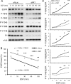
Biochemical analysis of EGFR phosphorylation. (A–C) Control shLuc and shR1 cells were exposed to 10 ng/ml EGF between 0 and 120 min, and the total EGFR and the phosphorylation (P) of specific tyrosine residues of EGFR were determined with specific antibodies as indicated. Reggie-1 (R1) and α-tubulin (α-tub) served as loading control. The quantification of the differences between shLuc and shR1 cells of total EGFR as well as each of the five tyrosine residues normalized to total EGFR are shown in B and C, respectively (n = 4, *p < 0.05, **p < 0.01, ***p < 0.001, paired t test).
FIGURE 6:
FIGURE 6:
Biochemical analysis of EGFR downstream targets. (A) shLuc and shR1 cells were stimulated with EGF as indicated in Figure 5. Western blot analyses show that the phosphorylation (P) of Src, PI3K subunits p85 and p55, Akt (T308 and S473), p38, and Erk1/2 kinases is altered in shR1 cells. Antibodies against total Src, PI3K subunit p85, Akt, p38, Erk1/2, reggie-1 (R1), and α-tubulin (α-tub) were used as loading control. (B) Quantification of the differences between shLuc and shR1 cells from A (n = 4, *p < 0.05, **p < 0 .01, **p < 0.001, paired t test).
FIGURE 7:
FIGURE 7:
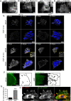
Formation of macropinosomes in A431 cells treated with EGF. (A) Reggie-1-EGFP (R1-EGFP)–transfected cells showed colocalization of reggie-1 and EGF-rhodamine (EGF-rhod) in the forming macropinosome after 5 min of EGF treatment (boxed area enlarged in inserts). (B) R1-EGFP colocalized with EGFR in a macropinosome. (C, D) Immunostaining with antibodies against E-cadherin (E-cad) and EGFR, as well as with antibodies against PrP and EGFR, showed colocalization of the protein pairs in macropinosomes. Scale bars, 10 μm. (E, F) Addition of Alexa-dextran (dextran) showed a significantly higher amount of internalization in shR1 cells compared with control shLuc cells (n = 3, ***p < 0.001, t test, mean ± SEM). Scale bars, 10 μm.
FIGURE 8:
FIGURE 8:
Macropinocytosis blockers rescue AJ formation in reggie-knockdown cells. (A–D) E-cadherin-EGFP (E-cad-EGFP)–transfected shR1 cells showed a recovery of AJ formation when treated with the macropinocytosis blockers amiloride (A), Rac1 (B), and PI3K (LY294002, D) but not when treated with the PLC blocker U-73122 (C). Selected areas showed cell contacts between E-cad–EGFP expressing and nontransfected cells for better visualization. Scale bars, 5 μm. (E) shLuc and shR1 cells were either nonstimulated (EGF 0 min) or stimulated with 10 ng/ml EGF for 20 min and immunostained with an antibody against the phosphorylated PI3K subunits p85 and p55 (P-PI3K). The weak staining of nonstimulated cells contrasts with the stronger signals after EGF stimulation, particularly in shLuc cells. Phosphorylated PI3K subunits accumulated at cell contacts in the apical pole of shR1 cells (boxed area), as opposed to a more widespread distribution in shLuc cells at apical and basal regions. Scale bars, 20 μm. (F) E-cad-EGFP–labeled vesicles of EGF-stimulated control shLuc and shR1 cells were monitored by time-lapse recordings at the focal plane of AJs for 1 min. The vesicle tracks were traced as indicated by the lines (right). Scale bars, 5 μm. (G) The quantification of E-cadherin vesicles emerging into the focal plane of AJs showed a significant increase in shR1 over control shLuc cells (n = 10, ***p < 0.001, t test, mean ± SEM). (H) E-cad-EGFP– and reggie-1-mRFP (R1-mRFP)– cotransfected A431 cells were stimulated with EGF for 15 min. A selected frame (boxed areas) showed that both proteins were colocalized at the same vesicles and tubulovesicular structures. Scale bars: 10 μm.
FIGURE 9:
FIGURE 9:
Model of the role of reggies and PrP in AJ formation. (A) In epithelial cells, PrP homophilic trans-interactions trigger clustering of PrP in reggie microdomains and activate Src-family tyrosine kinases at cell–cell contacts. This activated pool of Src kinases seems to be necessary for the correct phosphorylation of the Y845 residue of the EGFR during EGF stimulation. In addition, Y845 phosphorylation might be required for the proper autophosphorylation of various other tyrosine residues of the receptor, especially the Y1045 residue, which is known to be associated with the c-cbl–dependent EGFR internalization. EGF stimulation of EGFR at the PM, in turn, triggers the macropinocytosis of E-cadherin from AJs, which is recycled back to the cell contacts sites for the formation of new AJs. (B) The down-regulation of reggies or PrP generates a decrease in Src activation and an overall reduction in EGFR tyrosine phosphorylation during EGF stimulation. This impairs EGFR endocytosis, leading to the retention of the receptor at the PM and to the reduction in the activation of downstream molecules known to require EGFR-signaling endosomes. The increased surface EGFR signaling causes an enhanced macropinocytosis of E-cadherin from AJs. Thus the accelerated macropinocytosis and subsequent recycling of E-cadherin negatively affects the formation of AJs.

References

    1. Amaddii M, Meister M, Banning A, Tomasovic A, Mooz J, Rajalingam K, Tikkanen R. Flotillin-1/reggie-2 plays a dual role in the activation of receptor tyrosine kinase/map kinase signaling. J Biol Chem. 2012;287:7265–7278. - PMC - PubMed
    1. Araki N, Egami Y, Watanabe Y, Hatae T. Phosphoinositide metabolism during membrane ruffling and macropinosome formation in EGF-stimulated A431 cells. Exp Cell Res. 2007;313:1496–1507. - PubMed
    1. Baldys A, Gooz M, Morinelli TA, Lee MH, Raymond JR, Jr, Luttrell LM, Raymond JR., Sr Essential role of c-Cbl in amphiregulin-induced recycling and signaling of the endogenous epidermal growth factor receptor. Biochemistry. 2009;48:1462–1473. - PMC - PubMed
    1. Bryant DM, Kerr MC, Hammond LA, Joseph SR, Mostov KE, Teasdale RD, Stow JL. EGF induces macropinocytosis and SNX1-modulated recycling of E-cadherin. J Cell Sci. 2007;120:1818–1828. - PubMed
    1. Carcea I, Ma'ayan A, Mesias R, Sepulveda B, Salton SR, Benson DL. Flotillin-mediated endocytic events dictate cell type-specific responses to semaphorin 3A. J Neurosci. 2010;30:15317–15329. - PMC - PubMed

Publication types

MeSH terms