. 2012;7(3):e32661.
doi: 10.1371/journal.pone.0032661. Epub 2012 Mar 15.

The 3' untranslated regions of influenza genomic sequences are 5'PPP-independent ligands for RIG-I

Affiliations

The 3' untranslated regions of influenza genomic sequences are 5'PPP-independent ligands for RIG-I

William G Davis et al. PLoS One. 2012.

Abstract

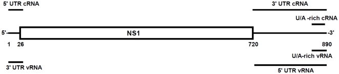
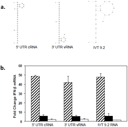
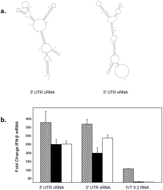
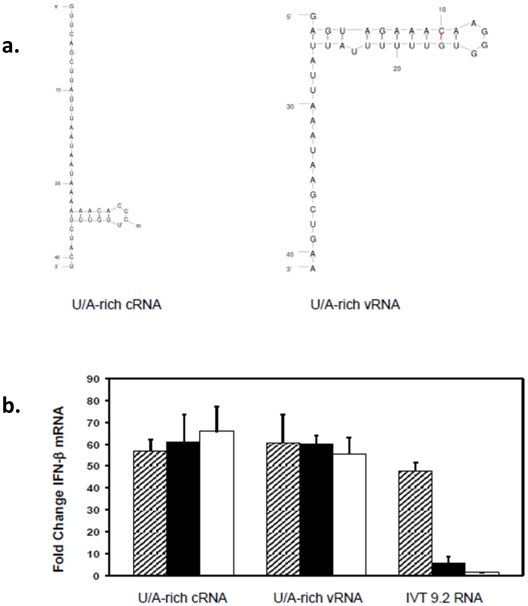
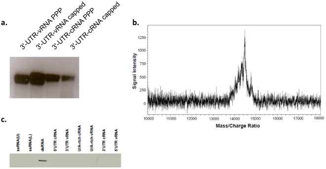
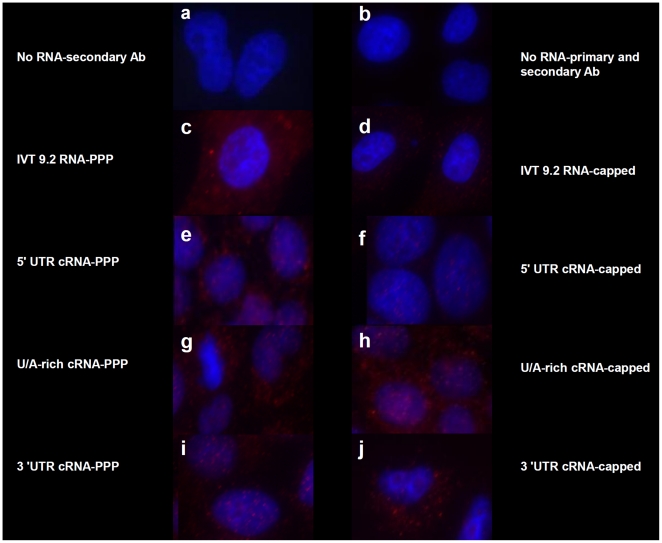
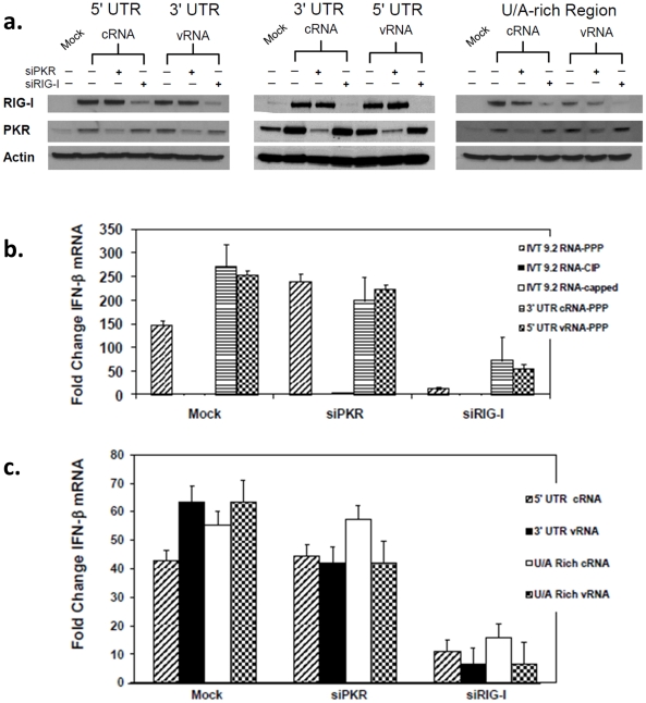
Retinoic acid inducible gene-I (RIG-I) is a key regulator of antiviral immunity. RIG-I is generally thought to be activated by ssRNA species containing a 5'-triphosphate (PPP) group or by unphosphorylated dsRNA up to ~300 bp in length. However, it is not yet clear how changes in the length, nucleotide sequence, secondary structure, and 5' end modification affect the abilities of these ligands to bind and activate RIG-I. To further investigate these parameters in the context of naturally occurring ligands, we examined RNA sequences derived from the 5' and 3' untranslated regions (UTR) of the influenza virus NS1 gene segment. As expected, RIG-I-dependent interferon-β (IFN-β) induction by sequences from the 5' UTR of the influenza cRNA or its complement (26 nt in length) required the presence of a 5'PPP group. In contrast, activation of RIG-I by the 3' UTR cRNA sequence or its complement (172 nt) exhibited only a partial 5'PPP-dependence, as capping the 5' end or treatment with CIP showed a modest reduction in RIG-I activation. Furthermore, induction of IFN-β by a smaller, U/A-rich region within the 3' UTR was completely 5'PPP-independent. Our findings demonstrated that RNA sequence, length, and secondary structure all contributed to whether or not the 5'PPP moiety is needed for interferon induction by RIG-I.


Conflict of interest statement

Competing Interests: The authors have declared that no competing interests exist.

Figures

Figure 1
Figure 1. Schematic representation of RNAs used in this study.
The influenza A virus segment 8 cRNA is shown with NS1 and NS2/NEP coding sequences boxed. The extended lines represent the 5′ and 3′ non-coding sequences. Bars (not drawn to scale) indicate sequences (see Materials and Methods) used to generate in vitro transcribed (IVT) RNAs.
Figure 2
Figure 2. 5′ PPP-independent induction of IFN-β by small influenza-derived RNA sequences.
(A) The secondary structures of the IVT RNAs were predicted using the program mfold (v3.2). (B) A549 cells were transfected with 3 µg of in vitro transcribed RNAs from the 5′ end of the cRNA/3′ end of the vRNA sequence of NS1 gene. 24 hr post-transfection, RNA was extracted to determine the levels of IFN-β by qRTPCR. The data are shown as folds over the mock control. Hatched bar, filled bar and empty bars represent untreated, CIP-treated and capped RNAs.
Figure 3
Figure 3. Induction of IFN-β message is triphosphate independent.
(A) The secondary structures of the IVT-RNAs shown were predicted by the program mfold (v3.2). (B) A549 cells were transfected with 3 µg of UTR RNA from the 3′ end of the cRNA or 5′ end of the vRNA and RNA was isolated 24 hr post-transfection to determine IFN-β levels by qRT-PCR. The data are shown as fold increases over levels in mock transfected cells. Error bars represent the standard deviation of triplicate qRT-PCR runs using RNAs from one of three representative experiments. Hatched bar, filled bar and empty bars represent untreated, CIP-treated and capped RNAs.
Figure 4
Figure 4. Smaller U/A rich IVT RNAs from the cRNA and vRNA UTRs are also triphosphate independent.
(A) The secondary structures of the IVT-RNAs used are presented. (B) A549 cells were transfected with U/A-rich cRNA and vRNA. 24 hours post-transfection, RNA was isolated to determine IFN-β mRNA levels by qRT-PCR. The data are shown as fold increases over levels in mock transfected cells. Hatched bar, filled bar and empty bars represent untreated, CIP-treated and capped RNAs. Error bars represent the standard deviation of triplicate qRT-PCR runs using RNAs from one of three representative experiments.
Figure 5
Figure 5. Homogeneity of IVT RNAs.
(A) Denaturing agarose gels of in vitro transcribed RNAs show products running as single bands. (B) U/A-rich vRNA was prepared by IVT and subjected to mass determination by MALDI-TOF mass spectroscopy. A single peak was observed spanning ∼1 kDa (13.8 k–14.8 k) and corresponding to the expected mass +/− ∼1–3 nucleotides. (C) To determine if the in vitro transcribed RNAs are ssRNA or ds NA, RNA samples were resolved on TAE PAGE, transferred onto nylon membrane and probed for dsRNA using dsRNA specific antibodies as described in Material and Methods. None of the in vitro transcribed RNAs nor the 41-nt long chemically synthesized ssRNA complementary strands were detected by the dsRNA-specific antibody. Only annealed 41 bp dsRNA was detected by the dsRNA-specific antibody.
Figure 6
Figure 6. 5′ PPP-independent activation of RIG-I.
A549 cells were transfected with 1 µg of the indicated IVT RNAs as in Figure 2. After 24 hrs, the cells were fixed with 4% paraformaldehyde and permeabilized with a 0.2% saponin/0.1% BSA/PBS buffer. Cells were blocked using CAS overnight and probed with a conformational dependent rabbit RIG-I polyclonal primary antibody that detects the RNA-bound form of RIG-I in the cytosol and followed by staining with AlexaFluor goat anti-rabbit 549 (stains red) and Hoechst 33342 (stains nucleus blue). Cells were visualized using a Zeiss fluorescent microscope with an axiocam HRM apotome attachment using AxioVision software.
Figure 7
Figure 7. Induction of IFN and IFN-stimulated genes is inhibited by reduction of RIG-I but not PKR.
A549cells were first transfected with the siRNAs against RIG-I or PKR(3 µg)as shown or mock transfected. 24 hr later, the cells were transfected again with the indicated IVT RNAs (3 µg) and processed 24 hr later for RNA as well as protein. Protein lysates are used to determine the levels of PKR and RIG-I proteins by western blot analyses and RNA is used to determine the levels of mRNA for IFN-β. (A) Western blots using the indicated antibodies of protein extracts from treated cells are shown. (B) and (C) IFN-β mRNA levels were measured using qRT-PCR. Error bars represent the standard deviation of triplicate qRT-PCR runs using RNAs from one of three representative experiments.

References

    1. Akira S. Innate immunity to pathogens: diversity in receptors for microbial recognition. Immunol Rev. 2009;227:5–8. - PubMed
    1. Seth RB, Sun L, Chen ZJ. Antiviral innate immunity pathways. Cell Res. 2006;16:141–147. - PubMed
    1. Diebold SS, Kaisho T, Hemmi H, Akira S, Reis e Sousa C. Innate antiviral responses by means of TLR7-mediated recognition of single-stranded RNA. Science. 2004;303:1529–1531. - PubMed
    1. Ranjan P, Jayashankar L, Deyde V, Zeng H, Davis WG, et al. 5′PPP-RNA induced RIG-I activation inhibits drug-resistant avian H5N1 as well as 1918 and 2009 pandemic influenza virus replication. Virol J. 2010;7:102. - PMC - PubMed
    1. Alexopoulou L, Holt AC, Medzhitov R, Flavell RA. Recognition of double-stranded RNA and activation of NF-kappaB by Toll-like receptor 3. Nature. 2001;413:732–738. - PubMed

MeSH terms