. 2012;7(3):e33138.
doi: 10.1371/journal.pone.0033138. Epub 2012 Mar 16.

A unique modification of the eukaryotic initiation factor 5A shows the presence of the complete hypusine pathway in Leishmania donovani

Affiliations

A unique modification of the eukaryotic initiation factor 5A shows the presence of the complete hypusine pathway in Leishmania donovani

Bhavna Chawla et al. PLoS One. 2012.

Abstract

Deoxyhypusine hydroxylase (DOHH) catalyzes the final step in the post-translational synthesis of an unusual amino acid hypusine (N(€)-(4-amino-2-hydroxybutyl) lysine), which is present on only one cellular protein, eukaryotic initiation factor 5A (eIF5A). We present here the molecular and structural basis of the function of DOHH from the protozoan parasite, Leishmania donovani, which causes visceral leishmaniasis. The L. donovani DOHH gene is 981 bp and encodes a putative polypeptide of 326 amino acids. DOHH is a HEAT-repeat protein with eight tandem repeats of α-helical pairs. Four conserved histidine-glutamate sequences have been identified that may act as metal coordination sites. A ~42 kDa recombinant protein with a His-tag was obtained by heterologous expression of DOHH in Escherichia coli. Purified recombinant DOHH effectively catalyzed the hydroxylation of the intermediate, eIF5A-deoxyhypusine (eIF5A-Dhp), in vitro. L. donovani DOHH (LdDOHH) showed ~40.6% sequence identity with its human homolog. The alignment of L. donovani DOHH with the human homolog shows that there are two significant insertions in the former, corresponding to the alignment positions 159-162 (four amino acid residues) and 174-183 (ten amino acid residues) which are present in the variable loop connecting the N- and C-terminal halves of the protein, the latter being present near the substrate binding site. Deletion of the ten-amino-acid-long insertion decreased LdDOHH activity to 14% of the wild type recombinant LdDOHH. Metal chelators like ciclopirox olamine (CPX) and mimosine significantly inhibited the growth of L. donovani and DOHH activity in vitro. These inhibitors were more effective against the parasite enzyme than the human enzyme. This report, for the first time, confirms the presence of a complete hypusine pathway in a kinetoplastid unlike eubacteria and archaea. The structural differences between the L. donovani DOHH and the human homolog may be exploited for structure based design of selective inhibitors against the parasite.


Conflict of interest statement

Competing Interests: The authors have declared that no competing interests exist.

Figures

Figure 1
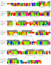
Figure 1. Comparison of the alignment and structural features of human (NP_112594) and L. donovani DOHH (ADJ39999) protein sequences with respect to the hypothetical protein YibA (NP_290174) of Escherichia coli.
This alignment gives an indication of structural features which would be expected for the L. donovani and human DOHH protein sequences. Residues in human and L. donovani DOHH are color-coded according to the chemical property of the amino acids. The number in parentheses after the protein code indicates the PDB residue number at the beginning of each block. The top line shows alignment positions. The gray block corresponds to residues in alpha helices. Key to the formatted Joy alignment of the E. coli DOHH (1OYZ) structure: Solvent inaccessible residues –UPPERCASE; Solvent accessible residues – lowercase; positive ϕ – italics; cis-peptide – breve ă; hydrogen bond to the other side chain – tilde ỹ; hydrogen bond to the main chain amide – bold; hydrogen bond to the main chain carbonyl – underlined; disulfide bond – cedilla ş.
Figure 2
Figure 2. Structure Modeling of DOHH.
(A) Model of the L. donovani DOHH protein generated by using the E. coli hypothetical protein YibA (Protein data bank accession ID: 1OYZ) as a template. As expected from the template structure, the generated model is rich in α-helices. The conserved His-Glu motifs are colored pink and are present at the inner concave surface of a horseshoe shaped structure. The amino acid insertions are colored yellow and are present at the outer surface and the inner concave surface of the generated model. (B) Superimposition of the L. donovani DOHH model (cyan) and the E. coli hypothetical protein YibA (yellow). The generated model and the E. coli structure superimpose with an RMSD of 1.6 Å. (C) Conservation pattern obtained for generated model of DOHH protein of L. donovani. DOHH is represented as a space-filled model and colored according to the conservation score. Fully conserved His-Glu motifs are marked by white arrows. The coloring scheme is depicted in the color-coding bar. All four His-Glu motifs are also represented. As is quite evident from the figure, the amino acid residues lining the inner concave surface are quite conserved as compared to the amino acid residues present on the outer surface. (D) Structural superposition human DOHH (violet) with that of L. donovani DOHH (Orange) is shown as a Ribbon diagram. The RMSD of the structural superposition is 4.8 Å. Metal chelating “HE” motifs from human (Pink) and L. donovani (Cyan) are shown as spheres. The sequence insertions in the L. donovani DOHH (white) are shown as cartoons.
Figure 3
Figure 3. Overexpression, Purification and Characterization of L. donovani DOHH protein.
(A) Overexpression of DOHH protein after induction at 3 hr with 0.5 mM IPTG. Lane 1, molecular weight marker (MBI Fermentas); Lanes 2 & 4, bacterial cell extract before induction; Lanes 3 & 5, bacterial cell extract after induction (B) Purification of DOHH protein on Ni2+-NTA acid affinity resin. Lane 1, molecular weight marker; Lane 2, Flowthrough; Lane 3-4, eluted fractions showing purified DOHH protein with buffer containing 150 mM imidazole. (C) Circular dichroism spectra of the recombinant DOHH showing that it is largely α-helical. The spectra were measured from 260 to 200 nm, at a bandwidth of 1 nm, using 100 µl of solution in a 0.1 mm path length cuvette and the analysis were performed as described in the Materials and Methods. (D) Comparison of the radioactivity in aminopropionaldehyde obtained as a result of [3H]hypusine formation after the DOHH assay with 1.0, 2.5, 5.0, 10.0 and 15.0 µg of the recombinant L. donovani DOHH protein. The reaction was performed as described in Materials and Methods. Results are mean ± SD of triplicate samples. (E) Comparison of radioactivity in aminopropionaldehyde obtained as a result of [3H]hypusine formation after the DOHH assay. The DOHH assay reactions were carried out as described in Materials and Methods. Recombinant DOHH enzyme, purified with and without 4 mM EDTA, was used in the absence and presence of 2 µM ferrous ammonium sulfate. The results are presented are the mean ± SD of triplicate samples. *, p<0.05, and ns indicates not significant (p>0.05).
Figure 4
Figure 4. Effect of deletion mutation on the activity of L. donovani DOHH.
(A) Scheme representing overlap extension PCR used for deletion of the ten-amino-acid-long insertion from LdDOHH. The regions shown in black and green are the regions upstream and downstream of the region to be deleted (shown in cyan blue). (B) Alignment showing human DOHH, LdDOHH and LdDOHH ▵(A169-V179). The regions that are similar between hDOHH and LdDOHH and align with each other are shown in red, yellow and blue color. Cyan blue color shows the insertions present in LdDOHH. (C) Comparison of the radioactivity in aminopropionaldehyde obtained as a result of [3H]hypusine formation after the DOHH assay with the recombinant LdDOHH enzyme or the mutant LdDOHH ▵(A169-V179) enzyme. The results presented are the mean ± SD of triplicate samples. *, p<0.05.
Figure 5
Figure 5. Effect of mimosine and CPX on deoxyhypusine hydroxylase activity.
Recombinant LdDOHH (5 µg) was incubated with either (A) mimosine (5, 10 or 20 µM) or (B) CPX (5, 10 and 15 µM) for 10 min at 37°C. Recombinant hDOHH was incubated with varying concentrations of (C) mimosine (20, 40, 60, 75 and 100 µM) and (D) CPX (15, 30, 45, 75 and 100 µM) for 10 min at 37°C. [3H]Hypusine formation was then measured as detailed in the Methods section. Results are mean ± SD of triplicate samples. *, p<0.05; **, p<0.002 and ns indicates not significant (p>0.05).

References

    1. Cooper HL, Park MH, Folk JE, Safer B, Braverman R. Identification of the hypusine-containing protein hy+ as translation initiation factor eIF-4D. Proc Natl Acad Sci U S A. 1983;80:1854–1857. - PMC - PubMed
    1. Park MH, Cooper HL, Folk JE. Identification of hypusine, an unusual amino acid, in a protein from human lymphocytes and of spermidine as its biosynthetic precursor. Proc Natl Acad Sci U S A. 1981;78:2869–2873. - PMC - PubMed
    1. Park MH, Cooper HL, Folk JE. The biosynthesis of protein-bound hypusine (N epsilon -(4-amino-2-hydroxybutyl)lysine). Lysine as the amino acid precursor and the intermediate role of deoxyhypusine (N epsilon -(4-aminobutyl)lysine). J Biol Chem. 1982;257:7217–7222. - PubMed
    1. Murphey RJ, Gerner EW. Hypusine formation in protein by a two-step process in cell lysates. J Biol Chem. 1987;262:15033–15036. - PubMed
    1. Chen KY, Dou QP. FEBS Lett 229: 325–328. 0014-5793(88)81149-9 [pii]; 1988. NAD+ stimulated the spermidine-dependent hypusine formation on the 18 kDa protein in cytosolic lysates derived from NB-15 mouse neuroblastoma cells. - PubMed

Publication types

MeSH terms

LinkOut - more resources