. 2012;7(3):e33609.
doi: 10.1371/journal.pone.0033609. Epub 2012 Mar 16.

TWEAK affects keratinocyte G2/M growth arrest and induces apoptosis through the translocation of the AIF protein to the nucleus

Affiliations

TWEAK affects keratinocyte G2/M growth arrest and induces apoptosis through the translocation of the AIF protein to the nucleus

Sanaa Sabour Alaoui et al. PLoS One. 2012.

Abstract

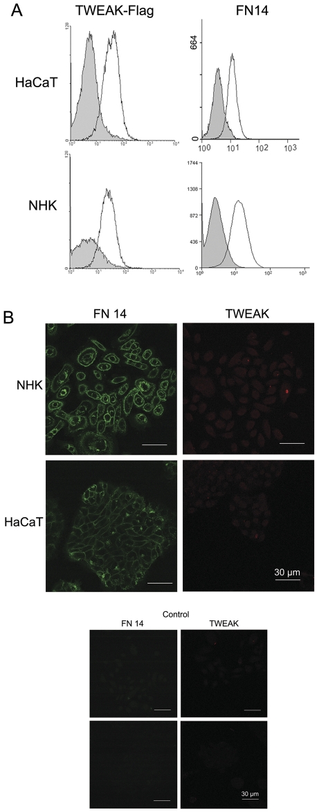
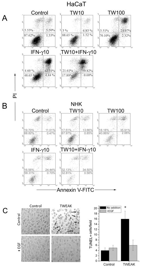
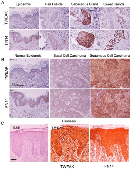
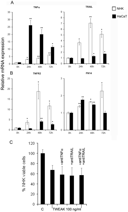
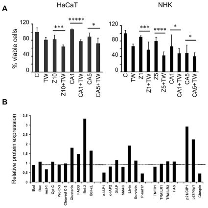
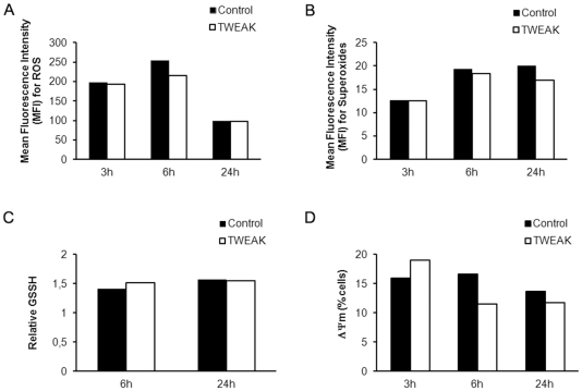
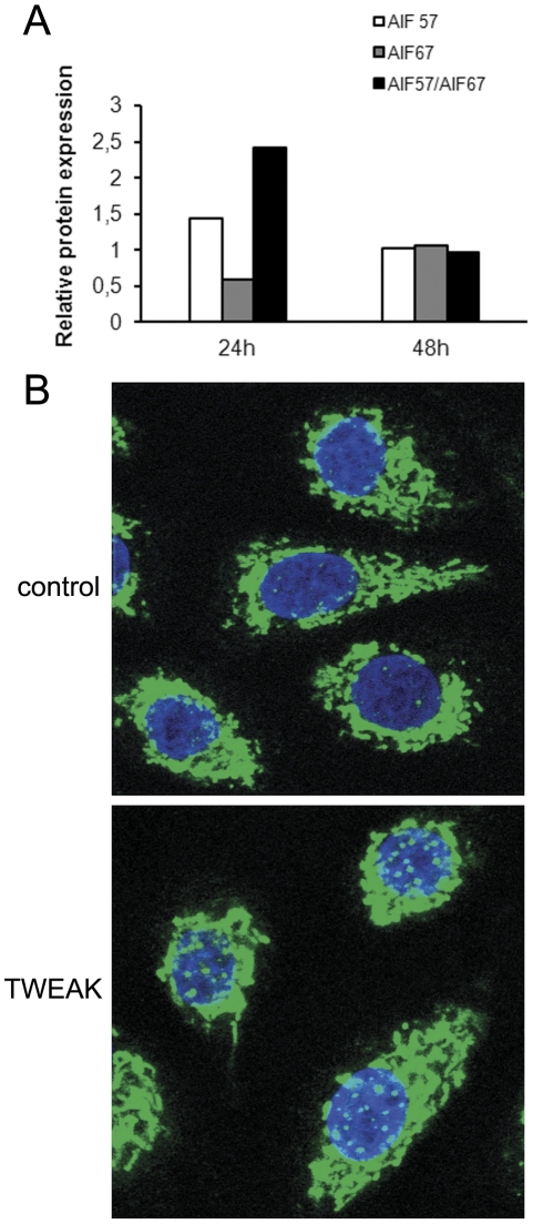
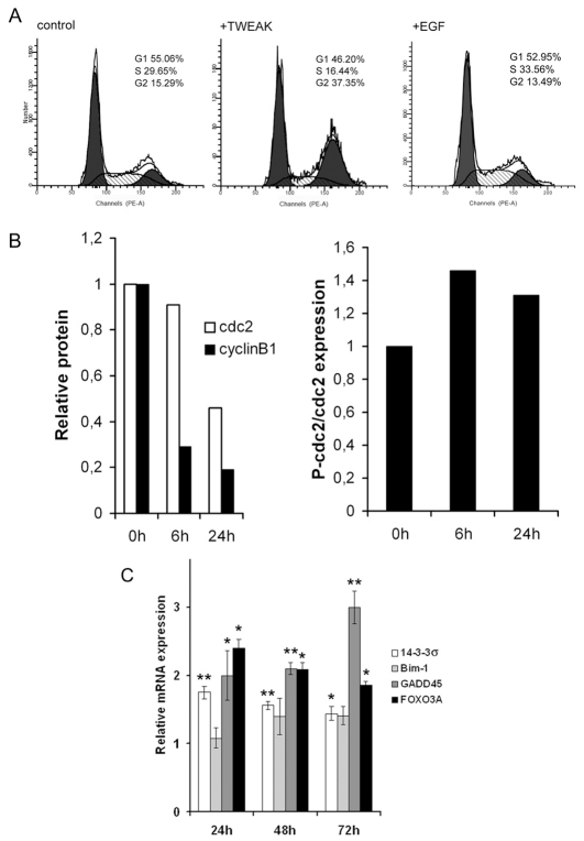
The soluble TNF-like weak inducer of apoptosis (TWEAK, TNFSF12) binds to the fibroblast growth factor-inducible 14 receptor (FN14, TNFRSF12A) on the cell membrane and induces multiple biological responses, such as proliferation, migration, differentiation, angiogenesis and apoptosis. Previous reports show that TWEAK, which does not contain a death domain in its cytoplasmic tail, induces the apoptosis of tumor cell lines through the induction of TNFα secretion. TWEAK induces apoptosis in human keratinocytes. Our experiments clearly demonstrate that TWEAK does not induce the secretion of TNFα or TRAIL proteins. The use of specific inhibitors and the absence of procaspase-3 cleavage suggest that the apoptosis of keratinocytes follows a caspase- and cathepsin B-independent pathway. Further investigation showed that TWEAK induces a decrease in the mitochondrial membrane potential of keratinocytes. Confocal microscopy showed that TWEAK induces the cleavage and the translocation of apoptosis inducing factor (AIF) from the mitochondria to the nucleus, thus initiating caspase-independent apoptosis. Moreover, TWEAK induces FOXO3 and GADD45 expression, cdc2 phosphorylation and cdc2 and cyclinB1 degradation, resulting in the arrest of cell growth at the G2/M phase. Finally, we report that TWEAK and FN14 are normally expressed in the basal layer of the physiological epidermis and are greatly enhanced in benign (psoriasis) and malignant (squamous cell carcinoma) skin pathologies that are characterized by an inflammatory component. TWEAK might play an essential role in skin homeostasis and pathology.


Conflict of interest statement

Competing Interests: The authors have declared that no competing interests exist.

Figures

Figure 1
Figure 1. FN14 is present on the cell surface and binds TWEAK.
A. The HaCaT cell line and normal human keratinocytes (NHK) were incubated in the presence of FLAG-tagged TWEAK and anti-human FN14 FITC-conjugated mAb. Control experiments were performed using cells incubated with buffer alone or in the presence of irrelevant mouse IgG FITC-conjugated antibodies. TWEAK binding was detected by the addition of the monoclonal M2 anti-FLAG antibody followed by the addition of a PE-conjugated anti-mouse IgG goat antibody. The grey area represents the mock control experiments, and the continuous line represents flagged-TWEAK, and anti-FN14 bound molecules. B. HaCaT cells and NHK were cytospun onto glass slides, were fixed and were stained using specific anti-FN14 FITC-conjugated and anti-TWEAK Texas red-conjugated mAbs. The slides were mounted and photographed as described in the Materials and Methods section.
Figure 2
Figure 2. TWEAK induces apoptosis of keratinocytes.
TWEAK induces the apoptosis of HaCaT cells (A) and NHKs (B) in a dose-dependent manner. HaCaT cells and NHKs were incubated in culture dishes with increasing amounts of TWEAK (10–100 ng/ml) and IFN-γ (10 ng/ml) for 48 h. The cells (including floaters) were collected and incubated with annexinV-FITC and propidium iodide for flow cytometry analysis. Control experiments were performed by incubating the cells in cell culture medium. The numbers indicate the percentage of cells within the indicated gate. The results shown are representative of three independent experiments. C. Normal human keratinocytes were examined under a microscope after the addition of recombinant TWEAK (100 ng/ml) or/and EGF (50 ng/ml) for 48 h. The characteristic membrane blebbing of the cells is shown by arrows, and quantification was performed using the TUNEL method. P values were calculated by comparing the treated cells to their relevant controls and were considered significant when P<0.05.
Figure 3
Figure 3. FN14 and TWEAK are detected in normal and pathological skin.
Representative sections of normal epidermis, hair follicles, and sebaceous and sweat glands (A), sections of epidermis from patients suffering from basal cell carcinoma, squamous cell carcinoma (B) and psoriasis (C) were stained for FN14 and TWEAK. Bar represents 200 µm.
Figure 4
Figure 4. Modification of the expression of death ligands and receptors in keratinocytes following the addition of TWEAK.
The relative expression of TNFα and TRAIL (A), TNFR2 and FN14(B) mRNAs in HaCaT cells and NHK compared with the expression of GAPDH mRNA. The results were normalized to the expression of control non-treated cells, which was given a value of 1. TWEAK was added at a concentration of 100 ng/ml. C. NHK were pre-incubated with neutralizing anti-TNFα (100 ng/ml) and anti-TRAIL (100 ng/ml) monoclonal antibodies before the addition of 100 ng/ml TWEAK. Cell viability in the presence and absence of neutralizing antibodies was measured 48 h later and is expressed as a percentage of the viability of control cells.
Figure 5
Figure 5. Apoptosis of keratinocytes is caspase- and cathepsin B-independent.
A. HaCaT cells (5.103 cells/well) or NHK (10.103 cells/well) were incubated with the indicated concentrations of caspase (zVAD-fmk) or cathepsin B (CA-074) inhibitors for 48 h (HaCaT) or 72 h (NHK) in the presence or absence of 100 ng/ml recombinant TWEAK. The cell viability was assessed using an MTT assay. The results are expressed as a percentage, with the proliferation of unstimulated cells set at 100% the (mean of triplicates ± SEM). The data are representative of three independent experiments. TWEAK and the inhibitors were not added to the control cells; however, DMSO at a concentration equal to that in the inhibitor solutions was added. Z = Zvad-fmk; C = CA-076; 1, 5 or 10 corresponds to 1, 5 or 10 µM, respectively; TW = TWEAK). P values were calculated by comparing the treated cells to the relevant controls, and were considered significant when P<0.05. B. NHK were treated with 100 ng/ml TWEAK for 24 h. The cells were lysed, and the expression of several pro- and anti-apoptotic proteins was measured as described in the Materials and Methods section. The relative expression of these proteins compared with that of cells not treated with TWEAK is presented. The controls are considered as 100% (dotted line).
Figure 6
Figure 6. TWEAK decreases the mitochondrial membrane potential of keratinocytes.
NHK were treated with TWEAK (100 ng/ml) and ROS (A), and superoxides (B), oxidized glutathione (C) and the mitochondrial membrane potential (C) was measured as indicated in the Materials and Methods section.
Figure 7
Figure 7. AIF translocates to the nucleus following the addition of TWEAK.
A. NHK cells were incubated with TWEAK (100 ng/ml). The cells were lysed at various time intervals and were analyzed by Western blotting with a rabbit polyclonal anti-AIF specific antibody. B. The control cells were left untreated. The cells were detached, placed on glass slides, fixed with paraformaldehyde, permeabilized with Triton X-100 and stained with a rabbit polyclonal anti-AIF antibody. The bands were visualized using donkey anti-rabbit F(ab)′2 FITC polyclonal antibody. The nucleus was stained with DAPI.
Figure 8
Figure 8. TWEAK induces the cell growth arrest of keratinocytes and the degradation of the cdc2/cyclin B1 complex, and activates FOXO3A, GADD45 and 14-3-3σ.
A. NHK cells were treated with TWEAK (100 ng/ml) or TWEAK and EGF (50 ng/ml) for 24 h. The nuclei were isolated, were stained using propidium iodide and the cells were counted using flow cytometry. The results were analyzed using a ModFit program. Untreated cells were used as controls. B. NHK cells were incubated with TWEAK (100 ng/ml). The cells were lysed at various time intervals and were analyzed by Western blotting with anti-cyclin B1, anti-cdc2 and anti-phospho-cdc2 specific antibodies. C. NHK cells were treated with TWEAK (100 ng/ml) for 24, 48 and 72 hours. The cells were lysed, and the RNA was isolated. A quantitative RT-PCR assay using specific primers for FOXO3A, GADD45, Bim-1 and 14-3-3σ mRNAs was used to measure the expression of these genes. GAPDH mRNA expression was used to normalize the results, which are expressed as a fold increment compared to that of the control sample, which was given a value of 1. A statistical analysis (the Student's t-test) was performed and the results were considered significant when P<0.05.

Similar articles

Cited by

References

    1. Chicheportiche Y, Bourdon PR, Xu H, Hsu YM, Scott H, et al. TWEAK, a new secreted ligand in the tumor necrosis factor family that weakly induces apoptosis. J Biol Chem. 1997;272:32401–32410. - PubMed
    1. Bodmer JL, Schneider P, Tschopp J. The molecular architecture of the TNF superfamily. Trends Biochem Sci. 2002;27:19–26. - PubMed
    1. Wiley SR, Cassiano L, Lofton T, Davis-Smith T, Winkles JA, et al. A novel TNF receptor family member binds TWEAK and is implicated in angiogenesis. Immunity. 2001;15:837–846. - PubMed
    1. Wiley SR, Winkles JA. TWEAK, a member of the TNF superfamily, is a multifunctional cytokine that binds the TweakR/Fn14 receptor. Cytokine Growth Factor Rev. 2003;14:241–249. - PubMed
    1. Browning JL, Miatkowski K, Sizing I, Griffiths D, Zafari M, et al. Signaling through the lymphotoxin beta receptor induces the death of some adenocarcinoma tumor lines. J Exp Med. 1996;183:867–878. - PMC - PubMed

Publication types

MeSH terms

Substances