. 2012;7(3):e33646.
doi: 10.1371/journal.pone.0033646. Epub 2012 Mar 16.

Immediate, but not delayed, microsurgical skull reconstruction exacerbates brain damage in experimental traumatic brain injury model

Affiliations

Immediate, but not delayed, microsurgical skull reconstruction exacerbates brain damage in experimental traumatic brain injury model

Loren E Glover et al. PLoS One. 2012.

Abstract

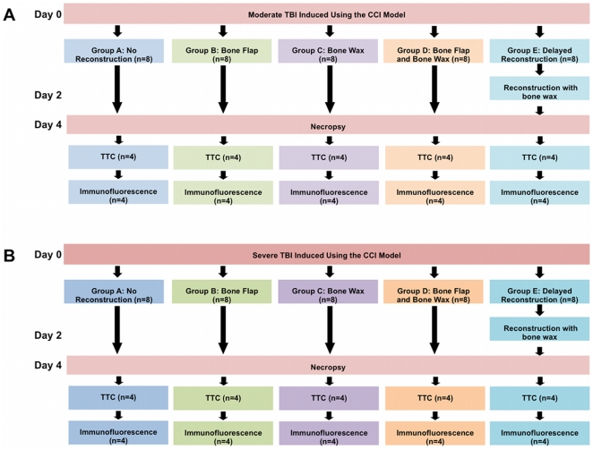
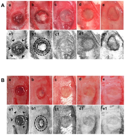
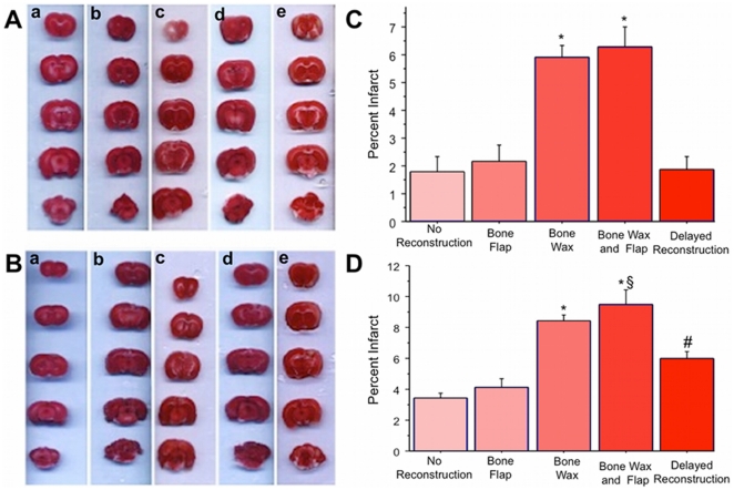
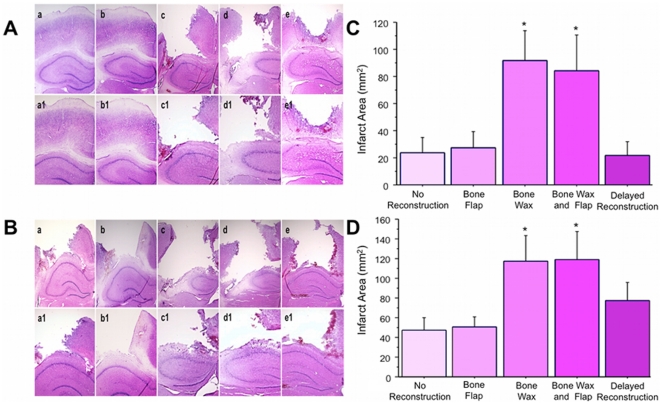
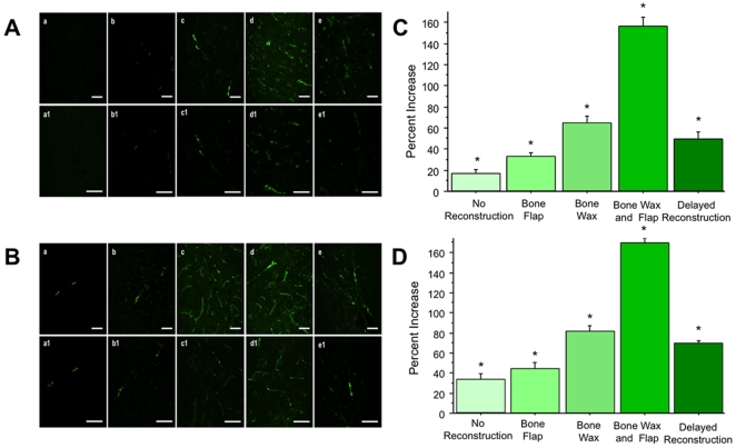
Moderate to severe traumatic brain injury (TBI) often results in malformations to the skull. Aesthetic surgical maneuvers may offer normalized skull structure, but inconsistent surgical closure of the skull area accompanies TBI. We examined whether wound closure by replacement of skull flap and bone wax would allow aesthetic reconstruction of the TBI-induced skull damage without causing any detrimental effects to the cortical tissue. Adult male Sprague-Dawley rats were subjected to TBI using the controlled cortical impact (CCI) injury model. Immediately after the TBI surgery, animals were randomly assigned to skull flap replacement with or without bone wax or no bone reconstruction, then were euthanized at five days post-TBI for pathological analyses. The skull reconstruction provided normalized gross bone architecture, but 2,3,5-triphenyltetrazolium chloride and hematoxylin and eosin staining results revealed larger cortical damage in these animals compared to those that underwent no surgical maneuver at all. Brain swelling accompanied TBI, especially the severe model, that could have relieved the intracranial pressure in those animals with no skull reconstruction. In contrast, the immediate skull reconstruction produced an upregulation of the edema marker aquaporin-4 staining, which likely prevented the therapeutic benefits of brain swelling and resulted in larger cortical infarcts. Interestingly, TBI animals introduced to a delay in skull reconstruction (i.e., 2 days post-TBI) showed significantly reduced edema and infarcts compared to those exposed to immediate skull reconstruction. That immediate, but not delayed, skull reconstruction may exacerbate TBI-induced cortical tissue damage warrants a careful consideration of aesthetic repair of the skull in TBI.


Conflict of interest statement

Competing Interests: Cesario V. Borlongan received funds from the following commercial entities: SanBio Inc., Celgene Cellular Therapeutics, KM Pharmaceutical Consulting, and NeuralStem Inc. There are no patents, products in development, or marketed products to declare. Cesario V. Borlongan is a PLoS ONE Editorial Board member. This does not alter the authors' adherence to all the PLoS ONE policies on sharing data and materials, as detailed online in the guide for authors.

Figures

Figure 1
Figure 1. Flowchart of experimental procedures.
All animals received moderate (Figure 1A) or severe (Figure 1B) TBI using the CCI model. After the CCI surgery, animals received either no reconstruction (Group A), immediate reconstruction with bone flap (Group B), bone wax (Group C), or bone wax and bone flap (Group D). The animals assigned to Group E in received delayed skull reconstruction on day 2. All animals were euthanized on day 4. Half of the animals (n = 4) from each group were assigned to TTC analysis and the other half (n = 4) were assigned to immunofluorescence assay.
Figure 2
Figure 2. Brain swelling accompanies TBI.
Following CCI, obvious brain swelling was detected in animals subjected to no skull reconstruction (Panels a and a1). Reconstruction with bone flap only provided partial skull reconstruction, thereby allowing a tempered brain swelling (Panels b and b1). Skull reconstruction with bone wax only (Panels c and c1), bone wax and bone flap (Panels d and d1), or delayed reconstruction with bone wax (Panels e and e1) afforded normalized skull structure, but also prevented visualization of brain swelling. Arrows are indicative of brain swelling following TBI. Dotted circles show the tempered brain swelling that occurred in the animals that received reconstruction with bone flap only.
Figure 3
Figure 3. TTC Immediate skull reconstruction with bone wax alone or in combination with bone flap exacerbates cortical damage in TBI.
TTC analysis of moderate (Figure 3A, quantified in C) and severe (Figure 3B, quantified in D) TBI revealed that immediate reconstruction with bone wax only or bone wax and bone flap significantly increased cortical damage compared to no reconstruction or reconstruction with only the bone flap. Of interest, delayed reconstruction at 2 days after TBI significantly reduced cortical damage compared to immediate reconstruction with bone wax only or bone wax and bone flap. Bars represent mean ± SEM. Asterisks (*) indicate p<0.05 vs. no reconstruction, immediate reconstruction with bone flap, and delayed reconstruction; # indicates p<0.05 vs. no reconstruction, bone flap, bone wax, and bone wax and flap; § indicates p<0.05 vs. bone wax.
Figure 4
Figure 4. Hematoxylin and eosin staining of moderate ( Figure 4A , with a–e at 2× magnification, a1–e1 at 4× magnification, and quantified in C) and severe ( Figure 4B , with a–e at 2× magnification, a1–e1 at 4× magnification, quantified in D) TBI models.
The cortical infarcts in the samples that received no reconstruction (Panel a, 2×; Panel a1, 4×) and immediate skull reconstruction with the bone flap only (Panel b, 2×; Panel b1, 4×) were smaller than the infarcts observed in the samples that received skull reconstruction with bone wax only (Panel c, 2×; Panel c1, 4×) and bone wax and bone flap (Panel d, 2×; Panel d1, 4×). The infarcts in the delayed reconstruction group (Panel e, 2×; Panel e1, 4×) were significantly reduced in both moderate and severe TBI models compared to the immediate reconstruction groups that received bone wax or bone wax and bone flap. Bars represent mean ± SEM. Asterisks * indicate p<0.05 vs. no reconstruction, bone flap, and delayed reconstruction.
Figure 5
Figure 5. Aquaporin-4 immunofluorescence of moderate ( Figure 5A
with a–e at 2× magnification, a1–e1 at 4× magnification, and quantified in C) and severe ( Figure 5B with a–e at 2× magnification, a1–e1 at 4× magnification, and quantified in C) TBI brains. Sparse aquaporin-4 staining was detected in the cortical tissues from TBI animals that received no reconstruction (Panel a, 40×; Panel a1, 60×). A slight, but significant increase in aquaporin-4 staining was seen in immediate skull reconstruction with bone flap (Panel b, 40×; Panel b1, 60×). Widespread aquaporin-4 upregulation was detected in the samples that received immediate skull reconstruction with bone wax only (Panel c, 40×; Panel c1, 60×) and bone wax and bone flap (Panel d, 40×; Panel d1, 60×), which was significantly elevated compared to no skull reconstruction. Aquaporin-4 density was reduced in the samples that received delayed reconstruction compared to immediate reconstruction with bone wax or bone wax and bone flap, but was significantly elevated compared to bone flap only or no reconstruction (Panel e, 40×; Panel e1, 60×). Bars represent mean ± SEM. Asterisks * indicate p<0.05 vs. all other treatment groups. Scale bars represent 50 µm.

References

    1. Faul M, Xu L, Wald M, Coronado V. Traumatic brain injury in the United States: emergency department visits, hospitalizations, and deaths. Atlanta (GA): Centers for Disease Control and Prevention. National Center for Injury Prevention and Control; 2010.
    1. Mammis A, McIntosh TK, Maniker AH. Erythropoietin as a neuroprotective agent in traumatic brain injury:: Review. Surgical neurology. 2009;71:527–531. - PubMed
    1. Yu S, Kaneko Y, Bae E, Stahl CE, Wang Y, et al. Severity of controlled cortical impact traumatic brain injury in rats and mice dictates degree of behavioral deficits. Brain research. 2009;1287:157–163. - PubMed
    1. Hayashi T, Kaneko Y, Yu SJ, Bae EK, Stahl CE, et al. Quantitative analyses of matrix metalloproteinase activity after traumatic brain injury in adult rats. Brain research. 2009;1280:172–177. - PubMed
    1. Bolkvadze T, Pitkanen A. Development of Post-Traumatic Epilepsy after Controlled Cortical Impact and Lateral Fluid-Percussion Induced Brain Injury in the Mouse. Journal of Neurotrauma 2011 - PubMed

Publication types

MeSH terms