. 2012 Mar 23:11:15.
doi: 10.1186/1476-4598-11-15.

c-Myb regulates matrix metalloproteinases 1/9, and cathepsin D: implications for matrix-dependent breast cancer cell invasion and metastasis

Affiliations

c-Myb regulates matrix metalloproteinases 1/9, and cathepsin D: implications for matrix-dependent breast cancer cell invasion and metastasis

Lucia Knopfová et al. Mol Cancer. .

Abstract

Background: The c-Myb transcription factor is essential for the maintenance of stem-progenitor cells in bone marrow, colon epithelia, and neurogenic niches. c-Myb malfunction contributes to several types of malignancies including breast cancer. However, the function of c-Myb in the metastatic spread of breast tumors remains unexplored. In this study, we report a novel role of c-Myb in the control of specific proteases that regulate the matrix-dependent invasion of breast cancer cells.

Results: Ectopically expressed c-Myb enhanced migration and ability of human MDA-MB-231 and mouse 4T1 mammary cancer cells to invade Matrigel but not the collagen I matrix in vitro. c-Myb strongly increased the expression/activity of cathepsin D and matrix metalloproteinase (MMP) 9 and significantly downregulated MMP1. The gene coding for cathepsin D was suggested as the c-Myb-responsive gene and downstream effector of the migration-promoting function of c-Myb. Finally, we demonstrated that c-Myb delayed the growth of mammary tumors in BALB/c mice and affected the metastatic potential of breast cancer cells in an organ-specific manner.

Conclusions: This study identified c-Myb as a matrix-dependent regulator of invasive behavior of breast cancer cells.


Figures
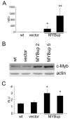
Figure 1
Figure 1
Derivation of MDA-MB-231 cells overexpressing c-myb. The MDA-MB-231 cells were transfected with human c-myb cDNA (MYBup) or the myb-less vector (vector) by lipofection. Pools of G418-resistant cells were cloned. (A) c-myb expression was determined in two independent clones by qRT-PCR. GAPDH was used as an internal control. (B) The c-Myb protein in two independent MYBup clones was determined by immunoblotting, and β-actin was used as a loading control. (C) Transactivation by c-Myb was determined by a luciferase assay using p6MBSluc as a reporter. The luciferase activity of each sample was expressed in relative light units and normalized for transfection efficiency according to the β-galactosidase activity. The columns show the average relative luciferase activity from three independent experiments. Error bars indicate standard deviations. Marks * and ** indicate significant (p < 0.05 and p < 0.01, respectively) differences in the relative amounts of c-myb mRNA (A) and in the luciferase activity (C) in the myb-less vector-transfected cells and MYBup cells as determined by the t-test.
Figure 2
Figure 2
Overexpression of c-Myb increases the migration and invasiveness of MDA-MB-231 cells through the basement membrane extract/Matrigel. (A) The transmigration assay was performed using Cultrex Cell Invasion Assay. MDA-MB-231MYBup and control cells migrating through a microporous membrane towards the chemoattractant (10% FCS)-containing medium were stained with the Calcein-AM. Cell migration was quantified according to the emitted fluorescence using the Synergy HT microplate reader. The columns show the average values of fluorescence from three independent measurements. (B) To determine cell invasiveness, the membrane was covered with a layer of the basement membrane matrix and the experiment was performed as described above. Error bars indicate the standard deviations. Asterisks indicate significant (p < 0.05) differences in the migration (A) and invasion (B) of the myb-less vector-transfected and MYBup cells as determined by the t-test.
Figure 3
Figure 3
Kinetics of c-Myb-induced migration and Matrigel invasion of MDA-MB-231MYBup cells. Cell migration/invasion in real time was analyzed by the xCELLigence RTCA. The control MDA-MB-231 (wt, vector), and the c-myb-transfected (M2, M5) cells were seeded at a density of 7.5 × 10⁴ per well and cultured in the chambers of CIM-plates. For the cell migration experiments, the membranes were left uncoated (A, B). For monitoring cell invasion, the membranes were coated with either Matrigel (C, D) or collagen I (E, F) as described in the Material and Methods. The bottom chambers were filled with either complete medium containing 10% FCS as a chemoattractant or with serum-free medium (-) in control wells. The charts show the representative outcomes of kinetics analysis of cell migration (A), Matrigel invasion (C) and collagen I invasion (E). The panels (B, D, F) show the average cell indexes at certain time points (6 h for migration and 12 h for invasion) from seven (migration) and five (invasion) independent measurements. Error bars indicate standard deviations. Asterisks indicate significant (p < 0.05) differences in the migration (B) and Matrigel invasion (D) of the myb-less vector-transfected cells and MYBup cells as determined by the t-test.
Figure 4
Figure 4
c-Myb upregulates cathepsin D and MMP9 and downregulates MMP1 in MDA-MB-231MYBup cells. (A) The relative amounts of MMP2, 3, 7, 10, 11, and 13 mRNA in the MYBup 5 and myb-less vector-transfected cells were determined by qRT-PCR. The relative amounts of MMP1, 9 and cathepsin D mRNAs were also determined in the MYBup 2 clone. Marks * and ** indicate significant (p < 0.05 and p < 0.01, respectively) differences in the relative amounts of cathepsin D, MMP9, and MMP1 mRNA in the myb-less vector-transfected and MYBup cells as determined by the t-test. The columns show the average values of relative normalized expression of the indicated genes from at least three independent experiments. (B) The control (wt, vector) and the MYBup cell protein extracts were analyzed by immunoblotting with anti-MMP1, anti-MMP9, and anti-cathepsin D antibodies. The secretion of cathepsin D, MMP9, and MMP1 was determined in the cell-conditioned medium. Equal amounts of total proteins were loaded. (C) To determine the activity of cathepsin D, protein extracts were prepared from harvested cells as described in the Material and Methods. The extent of hydrolysis of the fluorimetric substrate was measured in real time using a Synergy HT microplate reader. To demonstrate the assay specificity, pepstatin A was added to inhibit cathepsin D activity (data shown for MYBup 5 cells). (D) Transactivation assays were performed using six variants of luciferase reporters: two for cathepsin D (either containing the 2.85-kb or 0.82-kb promoter sequence), one for MMP9 (1.38 kb), and three for MMP1 (0.624, 1.606, and 4.3 kb). The luciferase activity of each sample was expressed in relative light units and normalized for transfection efficiency according to the β-galactosidase activity. The columns show the average values of relative luciferase activity from three independent measurements. Marks * and ** indicate significant (p < 0.05 and p < 0.01, respectively) differences in the luciferase activities in control cells and MYBup cells as determined by the t-test.
Figure 5
Figure 5
siRNA-mediated cathepsin D silencing reduces the migration and invasion of MDA-MB-231MYBup cells. MDA-MB-231MYBup cells were transfected with cathepsin D (CD) or control (ctrl) siRNA as described in the Material and Methods. (A) The level of cathepsin D protein in these cells was determined by immunoblotting. A representative western blot is presented. (B) Migration and invasion activities of the same cells were determined by the xCELLigence RTCA. The chart shows the representative outcomes of the kinetics analysis of cell migration. (C) The average cell indexes at the 6-h (migration, left) and 12-h (invasion, right) time points, respectively, from five independent measurements are shown. Error bars indicate standard deviations. Marks * and ** indicate significant (p < 0.05 and p < 0.01, respectively) differences in the migration/invasion of the cells transfected with cathepsin D siRNA and control siRNA as determined by the t-test.
Figure 6
Figure 6
c-Myb affects cell migration/invasion in murine 4T1 cells similarly as observed in MDA-MB-231 cells. 4T1 cells were transfected with murine c-myb cDNA (M-M5, M-M8) or with the myb-less vector (vector) by lipofection. G418-resistant cells were selected and cloned. (A) c-myb expression in both cell variants (vector and MYBup, respectively) and nontransfected cells (wt) was determined by immunoblotting. The 4T1MYBup variant was tested for migration (B) and invasion (C) activity using the xCELLigence RTCA as described in Figure 3. The panels represent the average cell indexes at certain time points (6 h for migration and 12 h for invasion) from three (collagen I) and four (migration and Matrigel invasion) independent measurements. Asterisks indicate significant (p < 0.05) differences in the migration (B) and Matrigel invasion (C) of the myb-less vector-transfected cells and MYBup cells as determined by the t-test. Error bars indicate standard deviations.
Figure 7
Figure 7
c-Myb delays mammary tumor growth and prevents the formation of pulmonary metastases. The tumor growth and spontaneous metastatic ability of the myb-less vector-transfected (control) 4T1 and 4T1M-M5 cell variant (MYBup) were determined following orthotopic inoculation into the mammary fat pads of BALB/c mice. (A) Tumor growth was monitored twice a week by measuring the tumor length (l) and width (w). Tumor volume was calculated using the equation l × w2/2. The mice injected with the MYBup 4T1 variants (n = 9) and control 4T1 cells (n = 9) were euthanized when the mean tumor diameter was approximately 1.2 cm. (B) Lungs were removed, rinsed in water, and fixed in Bouin's solution. Liver and bones were harvested and fixed in 10% buffered formalin. The number of lungs with surface metastases was determined using dissecting microscopy, and metastases in liver and bones were determined by histological examination of H&E-stained sections. Percentage of the mice with metastases (mts) developed in lung, liver and bones, respectively, is shown. (C) The surface metastatic nodules per lungs (mts) exemplified under the graph were enumerated by examination using dissecting microscopy. Asterisks indicate significant (p < 0.01) differences in the number of lung metastases in the mice injected with MYBup 4T1 variants and control 4T1 cells as determined by the t-test. Error bars indicate standard deviations. Representative samples of Bouin's solution-fixed tissues are shown. (D) Tissues were processed for paraffin embedding, sectioned, and stained with H&E. Bones were decalcified overnight before embedding. The lung, liver, and bone metastases were identified by light microscopy (arrows).

References

    1. Ramsay RG, Gonda TJ. MYB function in normal and cancer cells. Nat Rev Cancer. 2008;8:523–534. doi: 10.1038/nrc2439. - DOI - PubMed
    1. Roussel M, Saule S, Lagrou C, Rommens C, Beug H, Graf T, Stehelin D. Three new types of viral oncogene of cellular origin specific for haematopoietic cell transformation. Nature. 1979;281:452–455. doi: 10.1038/281452a0. - DOI - PubMed
    1. Jin S, Zhao H, Yi Y, Nakata Y, Kalota A, Gewirtz AM. c-Myb binds MLL through menin in human leukemia cells and is an important driver of MLL-associated leukemogenesis. J Clin Invest. 2010;120:593–606. doi: 10.1172/JCI38030. - DOI - PMC - PubMed
    1. Clappier E, Cuccuini W, Kalota A, Crinquette A, Cayuela JM, Dik WA, Langerak AW, Montpellier B, Nadel B, Walrafen P. et al.The C-MYB locus is involved in chromosomal translocation and genomic duplications in human T-cell acute leukemia (T-ALL), the translocation defining a new T-ALL subtype in very young children. Blood. 2007;110:1251–1261. doi: 10.1182/blood-2006-12-064683. - DOI - PubMed
    1. Lahortiga I, De Keersmaecker K, Van Vlierberghe P, Graux C, Cauwelier B, Lambert F, Mentens N, Beverloo HB, Pieters R, Speleman F. et al.Duplication of the MYB oncogene in T cell acute lymphoblastic leukemia. Nat Genet. 2007;39:593–595. doi: 10.1038/ng2025. - DOI - PubMed

Publication types

MeSH terms