. 2012 Mar 20;21(3):430-46.
doi: 10.1016/j.ccr.2011.12.029.

Transient low doses of DNA-demethylating agents exert durable antitumor effects on hematological and epithelial tumor cells

Affiliations

Transient low doses of DNA-demethylating agents exert durable antitumor effects on hematological and epithelial tumor cells

Hsing-Chen Tsai et al. Cancer Cell. .

Abstract

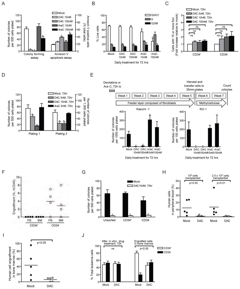
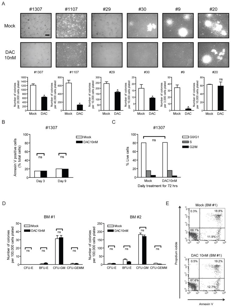
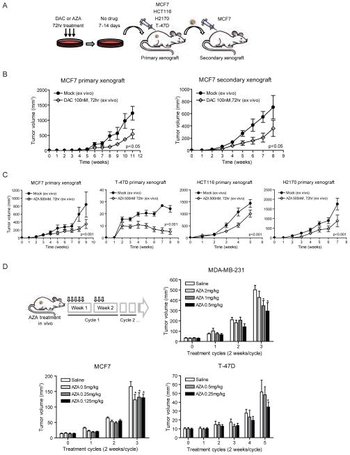
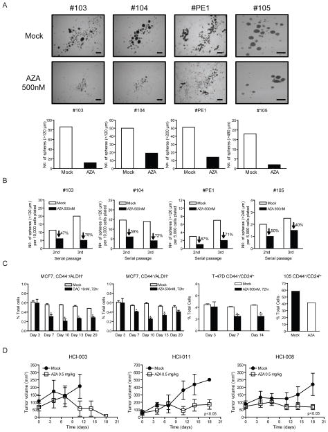
Reversal of promoter DNA hypermethylation and associated gene silencing is an attractive cancer therapy approach. The DNA methylation inhibitors decitabine and azacitidine are efficacious for hematological neoplasms at lower, less toxic, doses. Experimentally, high doses induce rapid DNA damage and cytotoxicity, which do not explain the prolonged time to response observed in patients. We show that transient exposure of cultured and primary leukemic and epithelial tumor cells to clinically relevant nanomolar doses, without causing immediate cytotoxicity, produce an antitumor "memory" response, including inhibition of subpopulations of cancer stem-like cells. These effects are accompanied by sustained decreases in genomewide promoter DNA methylation, gene reexpression, and antitumor changes in key cellular regulatory pathways. Low-dose decitabine and azacitidine may have broad applicability for cancer management.


Conflict of interest statement

Competing Interests: The authors declare no competing financial interests.

Figures

Figure 1
Figure 1. Low dose Decitabine (DAC) treatment diminishes self-renewing and leukemia-initiating capacities in cultured leukemia cells
(A) Apoptosis and methylcellulose colony forming assays of Kasumi-1 cells following 72-hour daily treatment with DAC or cytarabine (Ara-C). *p<0.05 compared to mock by ANOVA and Dunnett’s multiple comparison test. (B) Cell cycle analysis of Kasumi-1 after DAC or Ara-C daily treatment for 72 hours. *p<0.05 by ANOVA and Bonferroni post-tests. (C) γH2AX foci formation in CD34+ and CD34 Kasumi-1 cells after 72-hour DAC treatment. For each treatment, cells containing more than 15 foci are counted and calculated as fold change relative to that of mock treatment. ns, not statistically significant by ANOVA and Bonferroni post-tests. (D) Serial methylcellulose replating assays of Kasumi-1 after 72-hour daily treatment of DAC or Ara-C. Equal numbers of viable cells were plated for each plating *p<0.01 compared to mock by ANOVA and Bonferroni post-tests. (E) Long-term culture-initiating cell assay of Kasumi-1 and KG-1 AML cells following 72-hour daily treatment of DAC or Ara-C. *p<0.05 compared to mock by Mann-Whitney test. (F) Engraftment assay in NOD/Shi-scid/IL-2Rγnull mice of CD34+ versus CD34 Kasumi-1 cells. Human CD45+ cells were analyzed in the peripheral blood (PB) and Bone Marrow (BM) of transplant mice 4 to 5 months post transplantation. No engraftment was observed in any mice receiving CD34 cells. (G) Methylcellulose colony forming assay of CD34+ and CD34 Kasumi-1 cells following 72-hour daily treatment with 10nM DAC. *p<0.001, compared to mock, by ANOVA with Bonferroni posttests. (H and I) Engraftment assay in NOD/Shi-scid/IL-2Rγnull mice of Kasumi-1 cells pretreated daily with 10nM DAC for 72 hours. Percentage of human leukemia cells in peripheral blood (H) and in bone marrow (I) is shown. p values are calculated by Mann-Whitney test. (J) Percentage of CD34+ and CD34 Kasumi-1 AML cells after daily treatment of 10nM DAC for 72h in vitro and after engraftment in mouse bone marrow. *p<0.05 by Mann-Whitney test. All error bars represent standard errors. See also Figure S1.
Figure 2
Figure 2. Decitabine (DAC) at non-acutely-cytotoxic doses blunts clonogenicity of primary human leukemia cells but not normal bone marrow cells
(A) Methylcellulose colony forming assay of bone marrow mononuclear cells from 6 patients with newly diagnosed acute myelogenous leukemia (AML) following 72 hr daily DAC treatment and 4-day recovery period in vitro. Images and quantifications of colonies are shown in upper and lower panels, respectively. *p<0.05 compared to mock by ANOVA and Dunnett’s multiple comparison test. The scale bar represents 500μm. #1307 and #29: AML with FLT3-ITD mutation, #1107: AML FAB M2, #30: AML with mutated NPM1, #9: Secondary AML, #20: AML FAB M5. (B) Annexin V apoptosis assay of one representative AML patient sample (#1307) after 72hr daily DAC treatment in vitro. Bone marrow mononuclear cells were harvested for analysis at the end of 72 hr drug treatment (Day 3) and 6 days after drug removal (Day 9). ns, not statistically significant, by ANOVA with Bonferroni posttests. (C) Cell cycle analysis on one representative AML patient sample (#1307) was performed 4 days after drug removal (Day 7) with DNA content measured using propidium iodide staining. ns, not statistically significant, by ANOVA with Bonferroni posttests. (D) Methylcellulose colony forming assays of human normal bone marrow mononuclear cells (BM #1 and #2) following 72hr daily DAC treatment in vitro. CFU-E: Colony-Forming Unit-Erythroid, BFU-E: Burst-Forming Unit-Erythroid, CFU-GM: Colony-Forming Unit-Granulocyte, Macrophage, CFU-GEMM: Colony-Forming Unit-Granulocyte, Erythrocyte, Macrophage, Megakaryocyte. ns, not statistically significant, by ANOVA with Bonferroni posttests. (E) Annexin V apoptosis assay in one representative bone marrow sample (BM #1) after DAC treatment in vitro. Cells were double-stained with Annexin-V and propidium iodide, and percentage of cells relative to total cells is shown in each quadrant. All error bars represent standard errors.
Figure 3
Figure 3. Decitabine (DAC) and azacitidine (AZA) at non-cytotoxic doses decrease tumorigenicity in mouse tumor xenografts
(A) Schematic outline of xenograft tumorigenicity assay in NOD/SCID mice for MCF7 (breast), T-47D (breast), HCT116 (colon) and H2170 (lung) cancer cells after 7–14 day recovery period following 100 nM DAC or 500 nM AZA daily treatment for 72 hrs. (B) Primary and secondary xenograft assays of MCF7 cells pretreated with 100 nM DAC. Equal numbers of viable cells were injected in each transplantation. p value is calculated by ANOVA test. n=6 for each treatment group. (C) Primary xenograft assays of MCF7, T-47D, HCT116 and H2170 cells pretreated with 500 nM AZA. p value is calculated by ANOVA test. n=10 for each treatment group. (D) AZA treatment of mice with pre-established MDA-MD-231, MCF7 or T-47D breast tumors. Tumor volumes are measured at the end of each cycle. *Statistical significance is determined by two-way ANOVA test (p<0.05). All error bars represent standard errors. See also Figure S2.
Figure 4
Figure 4. Low dose Decitabine (DAC) and azacitidine (AZA) decrease self-renewal and tumorigenesis in primary breast cancer tissue from patients
(A) Tumor sphere assays of primary breast cancer cells from pleural effusions following 500nM AZA daily treatment for 3 days in vitro. Images and quantifications of tumor spheres are shown in upper and lower panels respectively. Scale bars represent 500μm. (B) Serial passages of tumor spheres formed by primary breast cancer cells following the initial 3-day AZA treatment. Equal numbers of viable cells were plated in each passage. The data shows a sustained decrease in self-renewal capacity of tumor spheres. (C) Flow cytometric analyses of CD44+/ALDH+ in MCF7 cells, CD44+/CD24lo cells in T-47D and primary breast cancer sample 105, following AZA or DAC daily treatment for 3 days and subsequent drug removal. Statistical significance was determined via two-way ANOVA test. (D) In vivo AZA treatment of immunodeficient mice bearing orthotopically-transplanted primary breast cancer cells from patients. 0.5mg/kg of AZA was administered intraperitoneally, 5 days a week. All error bars represent standard errors.
Figure 5
Figure 5. Low-dose decitabine (DAC) inhibits gene promoter DNA methylation, and causes sustained re-expression of hypermethylated genes
(A) Western blot of DNMT1 expression levels in human leukemia cells (Kasumi-1, KG-1a and KG-1) and breast cancer cells (MCF7, MDA-MB-231 and T-47D) following 72 hr daily treatment of 10 nM (leukemia) or 100 nM (breast cancer) DAC. (B) Methylation-Specific PCR (MSP) analyses of gene promoter DNA methylation in unsorted, CD34+ and CD34 Kasumi-1 cells prior to DAC treatment. U: unmethylated sequence amplifications, M: methylated sequence amplifications, IVD: in-vitro methylated DNA, NL: normal lymphocyte DNA. (C) MSP analyses of CDKN2B and CDH1 promoters in CD34+ and CD34 Kasumi-1 cells at 24, 48 and 72 hr of DAC treatment. (D) Quantitative real-time PCR analyses of CDKN2B gene expression over time in Kasumi-1 and KG-1a cells following 72 hr daily treatment of 10nM DAC. Expression levels are adjusted to GAPDH for each sample and graphed as fold changes relative to mock. All error bars represent standard errors.
Figure 6
Figure 6. Genome-wide methylation and expression analyses after short-term exporsure to low dose decitabine (DAC)
(A) Histogram of Infinium results for Kasumi-1 methylation profiles following daily DAC or ARA-C treatment for 72 hrs. Y-axis: frequency of probes. X-axis: CpG probe beta scores (lowest to highest = increasing DNA methylation). (B) Heat map composed of minimal variation beta probes located at −1,000 to +200 bp surrounding transcription start site (~ 5,500 genes). Blue and orange colors on the left denote non-CpG and CpG island promoters, respectively. PL6347, PL1, PL2, PL4, PL5: primary leukemia samples. PL6347+DAC: primary leukemia after 72hr 10 nM DAC treatment. mock and mock (rep): untreated Kasumi-1 cells and its replicate. Ara-C: Kasumi-1 cells treated with 100nM Ara-C daily for 72 hrs. TSA: Kasumi-1 cells treated with 300 nM trichostatin A for 9 hrs. DAC (day 3, day 3 rep, 7, 14, 21, 28): Kasumi-1 cells harvested at various time points post 72hr daily treatment of 10 nM DAC. (C) Box plots showing beta value changes over time in CpG island (upper panel) and non-CpG island gene promoters (lower panel) in Kasumi-1 cells following 72hr daily treatment of 10nM DAC. Ara-C: 100nM AraC treatment for 72 hrs. TSA: 300nM TSA treatment for 9 hrs. (D) Left panel: beta value changes after 72 hr, 10 nM DAC treatment for 6 genes in Kasumi-1 with basal beta values ≥ 0.5. Right panel: corresponding Agilent expression changes normalized to day 0. Gray horizontal lines: bottom to top, no change (log2 scale = 0), 1.4 fold (0.5) and 2.0 fold (1.0). See also Figure S3.
Figure 7
Figure 7. Metacore pathway analyses of Agilent expression changes following decitabine (DAC) or azacitidine (AZA) treatment
(A) Key pathways and/or master genes, shown here for studies in cultured cells. Genes in black = genes with expression changes not directly linked to promoter DNA methylation. Genes in red = genes with basal promoter hypermethylation, drug induced demethylation, and corresponding increases in expression. Superscripts by genes or pathways indicate: 1, Kasumi-1 AML cells, 2, KG-1a AML cells, and 3, MCF7 breast cancer cells with expression changes concordant in the indicated cell line. (B) An example of pathway diagram summarizes gene changes for cultured and primary AML and breast cancer samples after transient in vitro treatment of DAC or AZA. Events depicted illustrate key expression increases in cyclin dependent kinase inhibitors (CDKi’s – CDKN2B, CDKN2A and CDKN1A) which are known to trigger decreased activity of the FOXM1 pathway and all of the decreases shown above for FOXM1 itself and the other key participants for cycle entry and progression and including the oncogene, Skp2. (C) Analyses of DNA methylation by HumanMethylation450 BeadChip for multiple CpG sites in the CDKN1A proximal promoter region. Note that a normally heavily methylated (two normal bone marrow samples) region 5′ to the unmethylated CpG island (large green bar), surrounding an alternate gene start site (small arrow), is distinctly demethylated after 10 nM DAC treatment in 1107 primary AML cells which show all of the pathway changes but not to the same degree in 1307 AML cells where CDKN1A expression is not increased and the pathway changes seen are not present. See also Figure S4, Tables S1 and S2.
Figure 8
Figure 8. Validation of changes in multiple signaling pathways following decitabine (DAC) or azacitidine (AZA) treatment
(A) Quantitative real-time PCR verification for selected genes with expression changes indicated by the Metacore analyses. *p<0.05 determined by ANOVA and Bonferroni post-tests. (B) Quantitative real-time PCR analyses of gene expression changes in bone marrow samples taken from patients #1 (partial responder with AML) and #2 (complete responder with CMML) at pretreatment and various time points during or after administration of DAC (20mg/m2 IV over 1 hour daily × 5 days every 4 weeks). mRNA levels of two genes, CDKN2B and FOXM1, at each time point were ploted relative to the baseline level after adjusted by a control house-keeping gene, GAPDH. (C) Methylation-specific PCR analysis of CDKN2B promoter on the bone marrow sample of patient #1 at pretreatment, day 4, day 17 and day 30 during the treatment cycle. U: unmethylated sequence amplifications, M: methylated sequence amplifications. (D) Western blot analysis of total and phosphorylated AKT in MCF7 breast cancer cells post 100 nM DAC (upper panel) and 500 nM AZA (lower panel) treatment. (E) 3D assay in Matrigel of a primary breast cancer sample PE1 following 3 day treatment with 500 nM AZA and subsequent drug removal. Scale bars represent 100μm. (F) Flow cytometric analysis of surface CD11b expression in primary AML sample #29 at 14 days after 72 hr 10nM DAC treatment. (G) Immunofluoresence staining of cytokeratin 18 in cultured MCF7 cells 4 days after 72 hr 100nM DAC or 500 nM AZA daily treatment (upper panel), and in MCF7 xenograft tumors removed from mock and AZA-treated mice (lower panel) at 6 weeks. The scale bars represents 100μm. All error bars represent standard errors. See also Figure S5.

Comment in

References

    1. Abele R, Clavel M, Dodion P, Bruntsch U, Gundersen S, Smyth J, Renard J, van Glabbeke M, Pinedo HM. The EORTC Early Clinical Trials Cooperative Group experience with 5-aza-2′-deoxycytidine (NSC 127716) in patients with colo-rectal, head and neck, renal carcinomas and malignant melanomas. Eur J Cancer Clin Oncol. 1987;23:1921–1924. - PubMed
    1. Al-Hajj M, Wicha MS, Benito-Hernandez A, Morrison SJ, Clarke MF. Prospective identification of tumorigenic breast cancer cells. Proc Natl Acad Sci U S A. 2003;100:3983–3988. - PMC - PubMed
    1. Asou H, Tashiro S, Hamamoto K, Otsuji A, Kita K, Kamada N. Establishment of a human acute myeloid leukemia cell line (Kasumi-1) with 8;21 chromosome translocation. Blood. 1991;77:2031–2036. - PubMed
    1. Bachman KE, Rountree MR, Baylin SB. Dnmt3a and Dnmt3b are transcriptional repressors that exhibit unique localization properties to heterochromatin. J Biol Chem. 2001;276:32282–32287. - PubMed
    1. Berg T, Guo Y, Abdelkarim M, Fliegauf M, Lubbert M. Reversal of p15/INK4b hypermethylation in AML1/ETO-positive and -negative myeloid leukemia cell lines. Leuk Res. 2007;31:497–506. - PubMed

Publication types

MeSH terms

Associated data