. 2012 Jun;18(11-12):1213-28.
doi: 10.1089/ten.TEA.2011.0614. Epub 2012 May 10.

Further development of a tissue engineered muscle repair construct in vitro for enhanced functional recovery following implantation in vivo in a murine model of volumetric muscle loss injury

Affiliations

Further development of a tissue engineered muscle repair construct in vitro for enhanced functional recovery following implantation in vivo in a murine model of volumetric muscle loss injury

Benjamin T Corona et al. Tissue Eng Part A. 2012 Jun.

Abstract

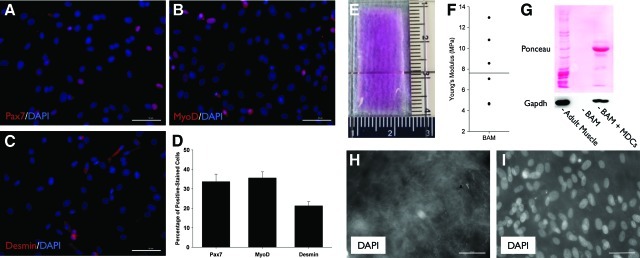
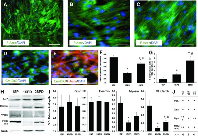
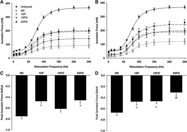
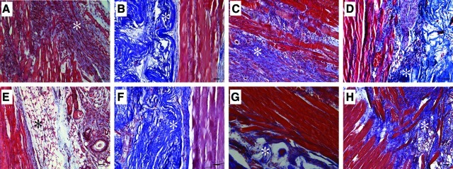
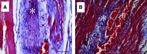
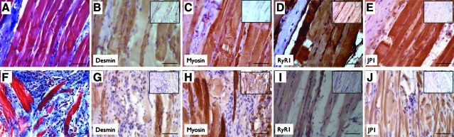
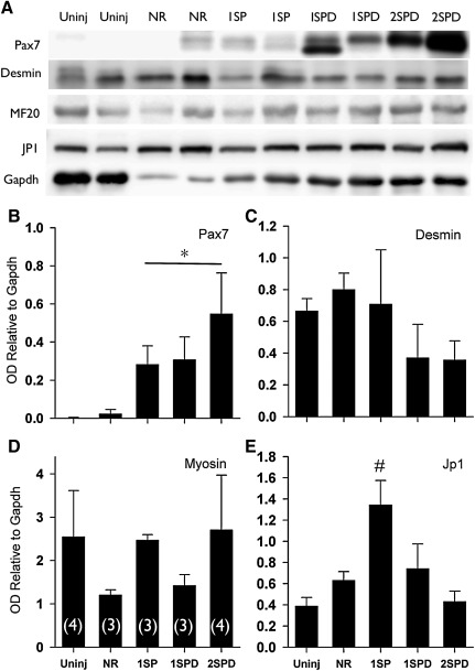
Volumetric muscle loss (VML) can result from trauma and surgery in civilian and military populations, resulting in irrecoverable functional and cosmetic deficits that cannot be effectively treated with current therapies. Previous work evaluated a bioreactor-based tissue engineering approach in which muscle derived cells (MDCs) were seeded onto bladder acellular matrices (BAM) and mechanically preconditioned. This first generation tissue engineered muscle repair (TEMR) construct exhibited a largely differentiated cellular morphology consisting primarily of myotubes, and moreover, significantly improved functional recovery within 2 months of implantation in a murine latissimus dorsi (LD) muscle with a surgically created VML injury. The present report extends these initial observations to further document the importance of the cellular phenotype and composition of the TEMR construct in vitro to the functional recovery observed following implantation in vivo. To this end, three distinct TEMR constructs were created by seeding MDCs onto BAM as follows: (1) a short-term cellular proliferation of MDCs to generate primarily myoblasts without bioreactor preconditioning (TEMR-1SP), (2) a prolonged cellular differentiation and maturation period that included bioreactor preconditioning (TEMR-1SPD; identical to the first generation TEMR construct), and (3) similar treatment as TEMR-1SPD but with a second application of MDCs during bioreactor preconditioning (TEMR-2SPD); simulating aspects of "exercise" in vitro. Assessment of maximal tetanic force generation on retrieved LD muscles in vitro revealed that TEMR-1SP and TEMR-1SPD constructs promoted either an accelerated (i.e., 1 month) or a prolonged (i.e., 2 month postinjury) functional recovery, respectively, of similar magnitude. Meanwhile, TEMR-2SPD constructs promoted both an accelerated and prolonged functional recovery, resulting in twice the magnitude of functional recovery of either TEMR-1SP or TEMR-1SPD constructs. Histological and molecular analyses indicated that TEMR constructs mediated functional recovery via regeneration of functional muscle fibers either at the interface of the construct and the native tissue or within the BAM scaffolding independent of the native tissue. Taken together these findings are encouraging for the further development and clinical application of TEMR constructs as a VML injury treatment.


Figures

FIG. 1.
FIG. 1.
Rat muscle derived cell (MDC) protein expression and bladder acellular matrix (BAM) scaffold characteristics. MDCs from primary culture were passaged once, seeded on noncoated chamber slides, and then cultured for 1 day in proliferation media (See Methods). Per protein marker (A–C), the total number of nuclei and positively stained nuclei were counted in at least 12 high-powered field (400×) images from at least two different chamber slides. Over 800 nuclei were counted for each protein marker with the number of positive cells expressed as percentage of total nuclei (D). BAM collagen scaffolds were cut to ∼3×1 sheet prior to implantation (E; scaffold was rehydrated in Dulbecco's Modified Eagle Medium for picture contrast). Young's modulus was determined for seven sterilized and rehydrated scaffolds (F). Scaffolds were confirmed to be decellularized via the absence of a protein (Ponceau) or specifically glyceraldehyde 3-phosphate Dehydrogenase (GAPDH) [blot; (G)], and the absence of nuclei [DAPI; (H)]. Protein expression (G) and nuclear staining via DAPI is demonstrated on BAM scaffold following the addition of MDCs (I). Scale bar=50 μm for all images. Color images available online at www.liebertonline.com/tea
FIG. 2.
FIG. 2.
Cellular morphology and protein expression characteristics of BAM-supported tissue engineered muscle repair (TEMR) constructs developed under three distinct culture conditions. TEMR-1SP, TEMR-1SPD, and TEMR-2SPD constructs are depicted in (A), (B), and (C), respectively (400× images). For the generation of the TEMR-2SPD constructs, a second batch of MDCs was added to an underlying layer of MDCs (i.e., TEMR-1SPD constructs). To confirm adherence of the second MDC batch, these cells were loaded with cytoplasmic fluorescent dye and then visualized following preconditioning (D and E). Scale bar=50 μm for all images. The number of nuclei (F) and the number of multinucleated cells (G) were quantitated for each construct type (See Methods, *TEMR-1SP; #TEMR-1SPD, p<0.05). Muscle-specific protein expression of TEMR constructs was characterized via Western blot (H). The optical densities of specified proteins were normalized to that of GAPDH for statistical comparisons among groups [(I); *significantly different from TEMR-1SP, p<0.05]. Protein expression of each construct type is summarized (J). Color images available online at www.liebertonline.com/tea
FIG. 3.
FIG. 3.
Latissimus dorsi (LD) muscle in vitro isometric force recovery following volumetric muscle loss (VML) injury is dependent on TEMR construct type. Uninjured and injured but nonrepaired (NR) or TEMR construct (three types, TEMR-1SP, TEMR-1SPD, and TEMR-2SPD)-repaired LD muscles were tested using direct muscle stimulation at 35°C in an organ bath (See Methods). Isometric force as a function of stimulation frequency was assessed for all experimental conditions at either 1 month (A) or 2 months (B) postinjury. Force–frequency curves were fit with a Hill equation as described in the Methods section. Peak isometric tetanic force functional deficits relative to the uninjured group mean was calculated for all experimental groups at 1 month (C) and 2 months (D). For each postinjury time, *to NR while #to all other groups (p<0.05). Values are expressed as means±standard error (SE). Sample sizes for each group at each postinjury time are listed in Table 1.
FIG. 4.
FIG. 4.
LD muscle tissue morphology after VML injury and immediate repair with TEMR constructs. VML-injured LD muscles that were either not repaired [(A) and (E)] or repaired with TEMR-1SP [(B) and (F)], TEMR-1SPD [(C) and (G)], or TEMR-2SPD [(D) and (H)] TEMR constructs were retrieved 1 month (A–D) and 2 months (E–H) postinjury and stained using Masson's trichrome (Red=tissue, Blue=Collagen, and Black=Nuclei). *Marker of area of initial injury [(A) and (E)] or presumptive BAM collagen deposition [(B–D) and (F–H)]. Images are 200× magnification with the scale bar=50 μm. Color images available online at www.liebertonline.com/tea
FIG. 5.
FIG. 5.
Presence of vascular and neural structures 1 month after TEMR construct treatment of VML-injured LD muscle. (A,B) Images are representative of vascular (#) and neural (*) structures that were identified via characteristic morphology and were observed in all TEMR construct groups 1 month postinjury. Images are 400× magnification; Scale bar=50 μm. Color images available online at www.liebertonline.com/tea
FIG. 6.
FIG. 6.
Functional protein expression in regenerating muscle fibers and putative neo-tissue 2 months after TEMR construct treatment of VML-injured LD muscle. Masson's trichrome staining and immunohistochemical staining for functional proteins is illustrated at the interface between the remaining native tissue and the TEMR construct [(A–E); representative of all TEMR repaired muscle] and for independent tissue formed in BAM scaffolding [(F–J); images derived from TEMR-1SPD and −2SPD-repaired muscle]. Insets show negative control staining for the primary antibody. Images are 400×magnification with the scale bar=50 μm. Color images available online at www.liebertonline.com/tea
FIG. 7.
FIG. 7.
LD muscle protein expression 2 months postinjury. (A) LD muscles were probed for Pax7, desmin, myosin (MF20), junctophilin 1 (JP1), and GAPDH using sodium dodecyl sulfate polyacrylamide gel electrophoresis and Western blotting. (B–E) Optical density was determined for each band and normalized to GAPDH. *Significantly different from uninjured; #Significantly different from all other groups (p<0.05). Values are expressed as mean±SE. Sample sizes for each group are listed in parentheses in panel D.

References

    1. Carlson B.M. Regeneration fo the completely excised gastrocnemius muscle in the frog and rat from minced muscle fragments. J Morphol. 1968;125:447. - PubMed
    1. Carlson B.M. Faulkner J.A. The regeneration of skeletal muscle fibers following injury: a review. Med Sci Sports Exerc. 1983;15:187. - PubMed
    1. Ciciliot S. Schiaffino S. Regeneration of mammalian skeletal muscle. Basic mechanisms and clinical implications. Curr Pharm Des. 2010;16:906. - PubMed
    1. Warren G.L. Summan M. Gao X. Chapman R. Hulderman T. Simeonova P.P. Mechanisms of skeletal muscle injury and repair revealed by gene expression studies in mouse models. J Physiol. 2007;582:825. - PMC - PubMed
    1. White T.P. Devor S.T. Skeletal muscle regeneration and plasticity of grafts. Exerc Sport Sci Rev. 1993;21:263. - PubMed

Publication types

MeSH terms

Substances