. 2012 Jun;23(6):1039-47.
doi: 10.1681/ASN.2011080776. Epub 2012 Mar 22.

Inducible podocyte injury and proteinuria in transgenic zebrafish

Affiliations

Inducible podocyte injury and proteinuria in transgenic zebrafish

Weibin Zhou et al. J Am Soc Nephrol. 2012 Jun.

Abstract

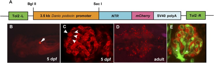
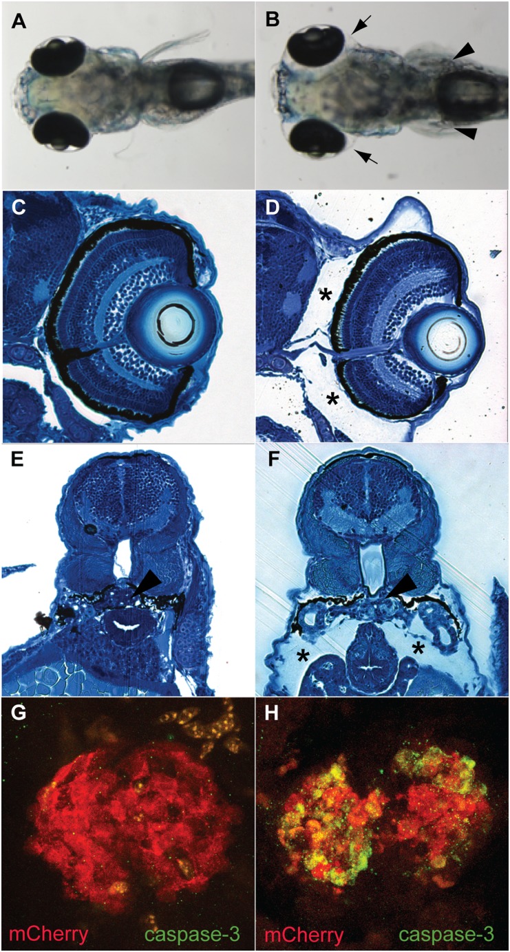
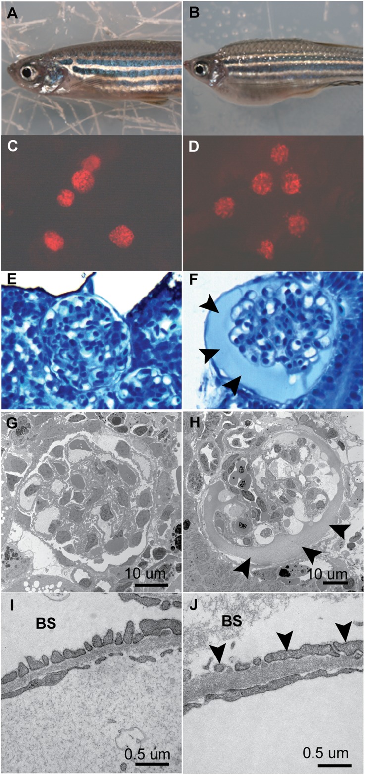
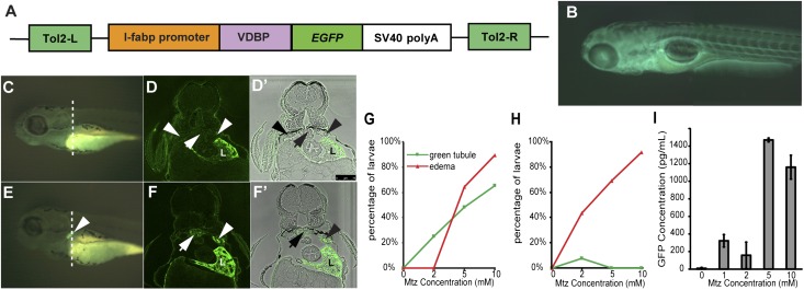
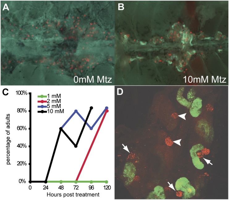
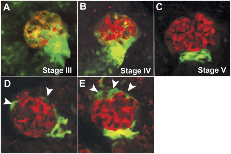
Damage or loss of podocytes causes glomerulosclerosis in murine models, and mutations in podocyte-specific genes cause nephrotic syndrome in humans. Zebrafish provide a valuable model for kidney research, but disruption of pronephroi leads to death within a few days, thereby preventing the study of CKD. In this study, we generated an inducible model of podocyte injury in zebrafish (pod::NTR-mCherry) by expressing a bacterial nitroreductase, which converts metronidazole to a cytotoxin, specifically in podocytes under the control of the zebrafish nphs2/podocin promoter. Application of the prodrug metronidazole to the transgenic fish induces acute damage to the podocytes in pronephroi of larval zebrafish and the mesonephroi of adult zebrafish, resulting in foot-process effacement and podocyte loss. We also developed a functional assay of the glomerular filtration barrier by creating transgenic zebrafish expressing green fluorescent protein (GFP)-tagged vitamin D-binding protein (VDBP) as a tracer for proteinuria. In the VDBP-GFP and pod::NTR-mCherry double-transgenic fish, induction of podocyte damage led to whole-body edema, and the proximal tubules reabsorbed and accumulated VDBP-GFP that leaked through the glomeruli, mimicking the phenotype of human nephrotic syndrome. Moreover, expression of wt1b::GFP, a marker for the developing nephron, extended into the Bowman capsule in response to podocyte injury, suggesting that zebrafish have a podocyte-specific repair process known to occur in mammalian metanephros. These data support the use of these transgenic zebrafish as a model system for studies of glomerular pathogenesis and podocyte regeneration.


Figures

Figure 1.
Figure 1.
Transgenic zebrafish expressing NTR-mCherry in pronephros and mesonephros. (A) Schematic graph illustrating the transgene structure of pod::NTR-mCherry. Tol2-L and Tol2-R are Tol2 transposon elements to facilitate the transgenesis. SV40 polyA is the polyadenylation signal sequence of simian virus 40. (B) Dorsal view of a 5-dpf larva. The pair of pronephric glomeruli (arrow) are marked with mCherry fluorescence (red). (C) Confocal image of pronephric glomerulus. The cell bodies (arrows) and major foot processes (arrow heads) of podocytes are visible by mCherry fluorescence. (D) Ventral view of medial nephron-dense region of mesonephros in adult zebrafish. Multiple mesonephric glomeruli (red) are labeled with mCherry fluorescence. (E) Confocal image of a mesonephric glomerulus in flk1::GFP/pod::NTR-mCherry double transgenic fish. Glomerular podocytes are red and endothelial cells are green. Branching of glomerular arterioles is notable (asterisks).
Figure 2.
Figure 2.
MTZ-induced podocyte injury in 5-dpf pod::NTR-mCherry transgenic fish. (A) pod::NTR-mCherry fish without MTZ treatment have normal morphologic features (dorsal view). (B) MTZ treatment of pod::NTR-mCherry fish leads to severe edema (arrowheads). (C and E) Cross-sections show normal morphologic features of untreated pod::NTR-mCherry transgenic zebrafish. (D and F) Note the periocular edema (asterisks) and edema around the body (asterisks). Arrowheads in E and F indicate the pronephric glomeruli. (G and H) Activated caspase-3 is detected in the pair of fused pronephric glomeruli in MTZ-treated pod::NTR-mCherry fish (H) but not in untreated wild-type fish (G).
Figure 3.
Figure 3.
MTZ-induced podocyte ablation leads to edema, glomerular leakage, and podocyte foot-process effacement in mesonephric glomeruli of adult pod::NTR-mCherry transgenic fish. (A and B) Induction of podocyte ablation by 24 hours of 10 mM MTZ in pod::NTR-mCherry fish causes severe edema. (C and D) Fluorescent images show the loss of pod::NTR-mCherry–expressing podocytes (red) in MTZ-treated mesonephric glomeruli. (E and F) Histologic sections reveal swelling of MTZ-treated mesonephric glomeruli and protein casts (arrowheads) in the Bowman space. (G and H) Electron microscopy reveals the swelling of glomeruli and a protein cast (arrowheads) in the Bowman space after MTZ treatment. (I and J) Higher-magnification images show podocyte foot-process effacement (arrowheads) in MTZ-treated fish. Note the protein cast in the Bowman space (BS) in MTZ-treated glomeruli.
Figure 4.
Figure 4.
Metronidazole-induced podocyte injury causes leakage of VDBP-GFP into proximal tubules in the pronephros of pod::NTR-mCherry/VDBP-GFP double-transgenic zebrafish larvae. (A) Schematic graph showing the l-fabp::VDBP-EGFP (enhanced green fluorescent protein) transgene. (B) Side view of 5-dpf l-fabp::VDBP-EGFP transgenic larvae. GFP fluorescence is evident in vasculature. (C) Side view of l-fabp::VDBP-GFP transgenic fish treated with 10 mM MTZ at 3–4 dpf shows no GFP accumulation in tubules (arrowhead). (D and D’). Cross-section of VDBP-GFP transgenic fish treated with 10 mM MTZ at the dash line in C stained with GFP antibody in fluorescence view (D) and merged with bright-field view (D’). Note GFP is absent in proximal tubules (arrowheads) and pronephric glomeruli (arrow). L, liver. Scale bar, 75 um. (E) Treatment with 10 mM MTZ on pod::NTR-mCherry/l-fabp::VDBP-GFP double transgenic fish at 3–4 dpf results in accumulation of VDBP-GFP in pronephric tubules (white arrowhead) due to glomerular leakage, compared with control (C). Note the reduction of GFP fluorescence in the head region of MTZ-treated fish (E) due to the loss of VDBP-GFP through damaged pronephros. (F and F’) Cross-section of MTZ-treated pod::NTR-mCherry/l-fabp::VDBP-GFP double transgenic fish at the dash line in E after GFP immunostaining in fluorescence view (F) and merged with bright-field view (F’) showing that GFP fluorescence is accumulated in proximal tubule (arrowheads) and pronephric glomeruli (arrow). L, liver. (G and H) Percentage of larvae manifesting VDBP-GFP accumulation and edema at 24 hours (G) and 48 hours (H) after MTZ treatment. (I) ELISA measurement of GFP in water after 24 hours of MTZ treatment on 3-dpf double transgenic fish.
Figure 5.
Figure 5.
MTZ-induced podocyte injury causes leakage of VDBP-GFP into proximal tubules in the mesonephros of pod::NTR-mCherry/VDBP-GFP double transgenic fish. (A) Ventral view of mesonephroi of adult double transgenic fish. (B) After 24 hours of 10 mM MTZ treatment, VDBP-GFP leaks through glomeruli and is reabsorbed in the proximal tubules. (C) MTZ-induced podocyte injury causing VDBP-GFP accumulation in proximal tubules in a dose-dependent manner. (D) Confocal image showing damaged mesonephroi and VDBP-GFP accumulation in proximal tubules. Note that severely damaged nephrons (arrows) exhibit VDBP-GFP accumulation in the proximal tubules, whereas the two nephrons (arrowheads) that are not severely damaged have little VDBP-GFP accumulation.
Figure 6.
Figure 6.
Postinjury response in zebrafish mesonephric glomeruli. (A–C) The distribution of wt1b::GFP-expressing cells (green) during the different developmental stages of glomeruli in adult zebrafish resembles that of renal progenitors in adult human glomeruli. With the maturation of the nephron, wt1b::GFP expression is diminished in the epithelial cells except at the urinary pole of the Bowman capsule. The podocytes (red) are labeled with mCherry fluorescence. (D and E) After MTZ-induced podocyte injury, wt1b::GFP-expressing cells (arrowheads) are expanded toward the vascular pole of the Bowman capsule of mesonephric glomeruli in zebrafish, suggesting a podocyte repair mechanism similar to that in mammals.

Comment in

References

    1. Kestilä M, Lenkkeri U, Männikkö M, Lamerdin J, McCready P, Putaala H, Ruotsalainen V, Morita T, Nissinen M, Herva R, Kashtan CE, Peltonen L, Holmberg C, Olsen A, Tryggvason K: Positionally cloned gene for a novel glomerular protein—nephrin—is mutated in congenital nephrotic syndrome. Mol Cell 1: 575–582, 1998 - PubMed
    1. Boute N, Gribouval O, Roselli S, Benessy F, Lee H, Fuchshuber A, Dahan K, Gubler MC, Niaudet P, Antignac C: NPHS2, encoding the glomerular protein podocin, is mutated in autosomal recessive steroid-resistant nephrotic syndrome. Nat Genet 24: 349–354, 2000 - PubMed
    1. Hasselbacher K, Wiggins RC, Matejas V, Hinkes BG, Mucha B, Hoskins BE, Ozaltin F, Nürnberg G, Becker C, Hangan D, Pohl M, Kuwertz-Bröking E, Griebel M, Schumacher V, Royer-Pokora B, Bakkaloglu A, Nürnberg P, Zenker M, Hildebrandt F: Recessive missense mutations in LAMB2 expand the clinical spectrum of LAMB2-associated disorders. Kidney Int 70: 1008–1012, 2006 - PubMed
    1. Zenker M, Aigner T, Wendler O, Tralau T, Müntefering H, Fenski R, Pitz S, Schumacher V, Royer-Pokora B, Wühl E, Cochat P, Bouvier R, Kraus C, Mark K, Madlon H, Dötsch J, Rascher W, Maruniak-Chudek I, Lennert T, Neumann LM, Reis A: Human laminin beta2 deficiency causes congenital nephrosis with mesangial sclerosis and distinct eye abnormalities. Hum Mol Genet 13: 2625–2632, 2004 - PubMed
    1. Löwik MM, Groenen PJ, Pronk I, Lilien MR, Goldschmeding R, Dijkman HB, Levtchenko EN, Monnens LA, van den Heuvel LP: Focal segmental glomerulosclerosis in a patient homozygous for a CD2AP mutation. Kidney Int 72: 1198–1203, 2007 - PubMed

Publication types

MeSH terms