Review
. 2012 Aug;1826(1):23-31.
doi: 10.1016/j.bbcan.2012.03.002. Epub 2012 Mar 13.

E-cadherin's dark side: possible role in tumor progression

Affiliations
Review

E-cadherin's dark side: possible role in tumor progression

Fausto J Rodriguez et al. Biochim Biophys Acta. 2012 Aug.

Abstract

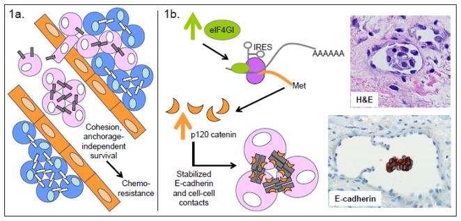
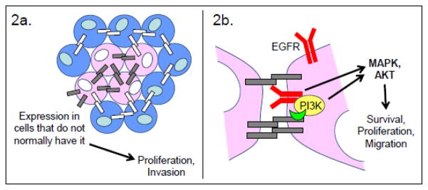
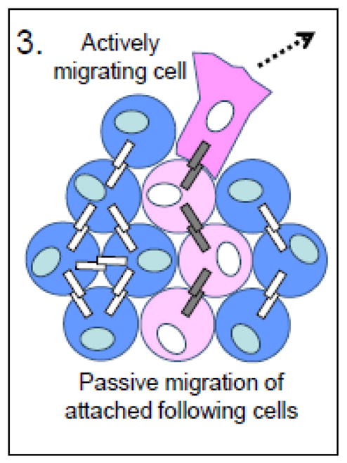
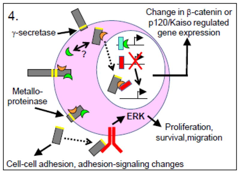
In the context of cancer, E-cadherin has traditionally been categorized as a tumor suppressor, given its essential role in the formation of proper intercellular junctions, and its downregulation in the process of epithelial-mesenchymal transition (EMT) in epithelial tumor progression. Germline or somatic mutations in the E-cadherin gene (CDH1) or downregulation by epigenetic mechanisms have been described in a small subset of epithelial cancers. However, recent evidence also points toward a promoting role of E-cadherin in several aspects of tumor progression. This includes preserved (or increased) E-cadherin expression in microemboli of inflammatory breast carcinoma, a possible "mesenchymal to epithelial transition" (MET) in ovarian carcinoma, collective cell invasion in some epithelial cancers, a recent association of E-cadherin expression with a more aggressive brain tumor subset, as well as the intriguing possibility of E-cadherin involvement in specific signaling networks in the cytoplasm and/or nucleus. In this review we address a lesser-known, positive role for E-cadherin in cancer.


Figures

Figure 1
Figure 1. Tumor-promoting functions of E-cadherin
Support of tumor cell survival, growth, and chemoresistance in the vasculature. a. In inflammatory breast carcinoma (IBC) models, E-cadherin is associated with the formation of intravascular tumor emboli. Experimentally, the increased cohesion mediated by E-cadherin leads to anchorage-independent survival of HSC-3 oral squamous cell carcinoma and nonepithelial Ewing tumor sarcoma cells and also promotes chemoresistance in Ewing tumor sarcoma and IBC cells. b. Experimental evidence from Silvera et al.[62] could explain the increased E-cadherin expression in IBC. Their data supports a model of IBC in which increased expression of the eIF4GI translation initiator leads to IRES-mediated translation of a specific mRNA molecular program that includes p120 catenin. The resulting increased expression of p120 catenin leads to stabilization of E-cadherin at cell junctions, formation and stabilization of tumor microemboli, and tumor dissemination in the lymphatics. H&E staining shows an IBC microembolus within the vasculature; E-cadherin staining shows its intense expression in the IBC microembolus. Blue cells represent normal cells; pink cells represent tumor cells; orange cells represent endothelial cells; gray bars represent E-cadherin; white bars represent any other type of cadherin.
Figure 2
Figure 2. Tumor-promoting functions of E-cadherin
Abnormal E-cadherin expression produces anomalous, E-cadherin-dependent activation of pro-survival, proliferative, and pro-migratory signaling. a. Aberrant expression of E-cadherin in cells that do not normally have it is a hallmark of ovarian carcinomas. This feature is also correlated with the increased proliferation and migration of a subset of glioblastoma tumor cells. b. Ligand-independent activation of the EGF receptor (EGFR) by E-cadherin has been demonstrated in ovarian and oral squamous cell carcinoma. Investigations with ovarian carcinoma indicate that the E-cadherin-catenin complex can coordinate the activation of PI3K, leading to localized pro-survival, pro-migratory AKT signaling. The E-cadherin-catenin complex can also coordinate the ErbB-family receptor tyrosine kinase-dependent activation of MAPK signaling, leading to proliferation. The precise molecular details of these interactions are not yet clear. Blue cells represent normal cells; pink cells represent tumor cells; orange cells represent endothelial cells; gray bars represent E-cadherin; white bars represent any other type of cadherin.
Figure 3
Figure 3. Tumor-promoting functions of E-cadherin
Facilitation of collective cell migration. Collective cell migration allows squamous cell carcinoma cells with intact E-cadherin-based junctions to move passively through a tissue by being pulled along by an actively migrating tumor cell. Blue cells represent normal cells; pink cells represent tumor cells; orange cells represent endothelial cells; gray bars represent E-cadherin; white bars represent any other type of cadherin.
Figure 4
Figure 4. Tumor-promoting functions of E-cadherin
Aberrant signaling due to proteolytic cleavage fragments of E-cadherin. (Green crescent = β-catenin; orange crescent = p120 catenin; blue rectangle = Lef-1/Tcf transcription factors; red rectangle = Kaiso transcription regulator.) Proteolytic cleavage of E-cadherin by either γ-secretases or metalloproteinases produces cleavage fragments that influence cell-cell adhesion as well as, respectively, gene transcription events or adhesion-related or growth factor receptor signaling. For example, the cytoplasmic portion of E-cadherin may serve on the one hand as a source/sink for β-catenin, thereby regulating β-catenin/Lef-1/Tcf transcriptional events, or on the other hand as a co-regulator with p120 catenin of Kaiso-mediated transcriptional repression. Likewise, the soluble extracellular fragment of E-cadherin may alter existing adherens junctions, or may stimulate signaling through EGFR or other growth factor receptors. Blue cells represent normal cells; pink cells represent tumor cells; orange cells represent endothelial cells; gray bars represent E-cadherin; white bars represent any other type of cadherin.

References

    1. Takeichi M. Cadherin cell adhesion receptors as a morphogenetic regulator. Science. 1991;251:1451–1455. - PubMed
    1. Nagafuchi A, Takeichi M. Cell binding function of E-cadherin is regulated by the cytoplasmic domain. EMBO J. 1988;7:3679–3684. - PMC - PubMed
    1. Perez-Moreno M, Jamora C, Fuchs E. Sticky business: orchestrating cellular signals at adherens junctions. Cell. 2003;112:535–548. - PubMed
    1. van Roy F, Berx G. The cell-cell adhesion molecule E-cadherin. Cell Mol Life Sci. 2008;65:3756–3788. - PMC - PubMed
    1. Cavallaro U, Christofori G. Cell adhesion and signalling by cadherins and Ig-CAMs in cancer. Nat Rev Cancer. 2004;4:118–132. - PubMed