Review
. 2012 Feb;2(1):43-9.
doi: 10.1016/j.coviro.2011.12.002. Epub 2011 Dec 23.

Targeted entry of enveloped viruses: measles and herpes simplex virus I

Affiliations
Review

Targeted entry of enveloped viruses: measles and herpes simplex virus I

Chanakha K Navaratnarajah et al. Curr Opin Virol. 2012 Feb.

Abstract

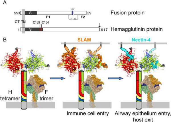
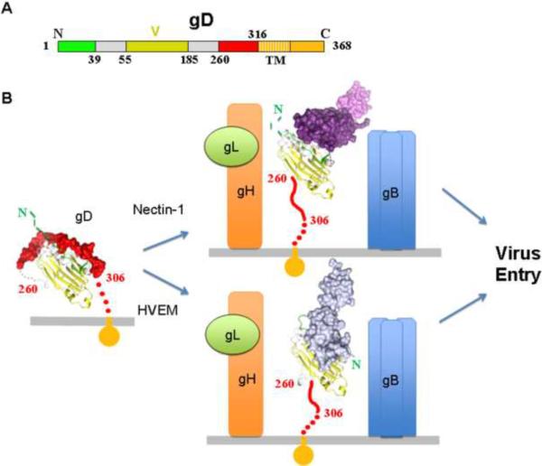
We compare the receptor-based mechanisms that a small RNA virus and a larger DNA virus have evolved to drive the fusion of viral and cellular membranes. Both systems rely on tight control over triggering the concerted refolding of a trimeric fusion protein. While measles virus entry depends on a receptor-binding protein and a fusion protein only, the herpes simplex virus (HSV) is more complex and requires four viral proteins. Nevertheless, in both viruses a receptor-binding protein is required for triggering the membrane fusion process. Moreover, specificity domains can be appended to these receptor-binding proteins to target virus entry to cells expressing a designated receptor. We discuss how principles established with measles and HSV can be applied to targeting other enveloped viruses, and alternatively how retargeted envelopes can be fitted on foreign capsids.


Figures

Figure 1
Figure 1
MV fusion complex. A. Schematic of MV F and H proteins indicating cytoplasmic tail (CT), transmembrane (TM), stalk (S) and globular head regions. For the F-protein the fusion peptide (FP, blue shading) and the F1 and F2 subunits that are the result of an activating proteolytic cleavage are indicated. The first residue of the F2 protein is His29; residues 1–28 constitute the signal peptide. The inter-subunit disulfide bond between F1 and F2 is indicated (s–s). The inter-dimer disulfide bonds in the H-dimer are indicated by red lines (C139 and C154). Targeting ligands are appended to the C-terminus of the H-protein as indicated by an arrow. B. Left panel. The H-tetramer is indicated as a combination of a schematic (stalk) and crystal structure[45] (two H-head dimers). Each monomer of the H-tetramer is shaded in a unique color which is extended to the stalk schematic. The C-terminal residue of the crystal structure is indicated by a grey shaded space-filling amino acid (residue 607). The MV F-trimer has been modeled based on the crystal structure of the related human paramyxovirus 5 F-trimer[46]. Each monomer has been shaded in a different color for clarity. The fusion peptide has been shaded blue. Middle panel. The SLAM-binding footprint has been shaded orange and a schematic used to depict the approximate SLAM-binding location. Right panel. The nectin4-binding footprint is indicated by cyan shading. The proposed location of nectin4-binding is indicated by a schematic. Each oval represents an Ig-like domain. Only two receptor molecules are drawn for simplicity, but up to four receptor molecules may bind one H-tetramer to trigger fusion.
Figure 2
Figure 2
Involvement of the HSV receptor-binding protein gD in entry. A. Schematic representation of HSV-1 gD. Numbering for gD starts at lysine 1 of the mature glycoprotein. The gD IgG core, labeled V for the Ig V-fold, is colored in yellow, residues forming the HVEM binding hairpin are in green and residues 260 to 316 that are displaced by receptor binding are in red. The gD Ig core has been shown to be replaceable when used for retargeting the virus. B. Structural schematic showing the triggering of the entry process mediated by gD. The same color code as in panel A was used. Regions that were not determined and presumably flexible in the crystal structures are shown as dotted lines. The left panel shows gD in the unliganded state where residues 260–306 are folded against the core structure. Nectin-1 or HVEM receptors bind to gD causing unwrapping of this C-terminal region (central panel). Exposure of this region mediates gH/gL (orange/green, respectively) and gB (blue trimer) interactions with concomitant membrane fusion and viral entry. The location of the C-terminal region of gD (residues 260–316) was not determined in the gD/Nectin-1 and gD/HVEM structures and is modeled as an extended coil pointing towards the viral membrane. Similarly the N-terminal 20 amino acids of gD were not localized in the unliganded gD and in the gD/Nectin-1 complex.

References

    1. Galanis E. Cancer: Tumour-fighting virus homes in. Nature. 2011;477:40–41. - PubMed
    1. Cattaneo R, Miest T, Shashkova EV, Barry MA. Reprogrammed viruses as cancer therapeutics: targeted, armed and shielded. Nat. Rev. Microbiol. 2008;6:529–540. - PMC - PubMed
    1. Ferreira CSA, Frenzke M, Leonard VHJ, Welstead GG, Richardson CD, Cattaneo R. Measles virus infection of alveolar macrophages and dendritic cells precedes spread to lymphatic organs in transgenic mice expressing human signaling lymphocytic activation molecule (SLAM, CD150) J. Virol. 2010;84:3033–3042. - PMC - PubMed
    1. Lemon K, De Vries RD, Mesman AW, Mcquaid S, Van Amerongen G, Yüksel S, Ludlow M, Rennick LJ, Kuiken T, Rima BK, et al. Early target cells of measles virus after aerosol infection of non-human primates. PLoS Pathogens. 2011;7:e1001263. - PMC - PubMed
    1. Ludlow M, Rennick LJ, Sarlang S, Skibinski G, McQuaid S, Moore T, de Swart RL, Duprex WP. Wild-type measles virus infection of primary epithelial cells occurs via the basolateral surface without syncytium formation or release of infectious virus. J. Gen. Virol. 2010;91:971–979. - PubMed

Publication types