Systematic approaches to central nervous system myelin
- PMID: 22441408
- PMCID: PMC11114939
- DOI: 10.1007/s00018-012-0958-9
Systematic approaches to central nervous system myelin
Abstract
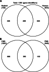
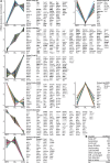
Rapid signal propagation along vertebrate axons is facilitated by their insulation with myelin, a plasma membrane specialization of glial cells. The recent application of 'omics' approaches to the myelinating cells of the central nervous system, oligodendrocytes, revealed their mRNA signatures, enhanced our understanding of how myelination is regulated, and established that the protein composition of myelin is much more complex than previously thought. This review provides a meta-analysis of the > 1,200 proteins thus far identified by mass spectrometry in biochemically purified central nervous system myelin. Contaminating proteins are surprisingly infrequent according to bioinformatic prediction of subcellular localization and comparison with the transcriptional profile of oligodendrocytes. The integration of datasets also allowed the subcategorization of the myelin proteome into functional groups comprising genes that are coregulated during oligodendroglial differentiation. An unexpectedly large number of myelin-related genes cause-when mutated in humans-hereditary diseases affecting the physiology of the white matter. Systematic approaches to oligodendrocytes and myelin thus provide valuable resources for the molecular dissection of developmental myelination, glia-axonal interactions, leukodystrophies, and demyelinating diseases.
Figures


Similar articles
-
Intra- and intercellular trafficking in sphingolipid metabolism in myelination.Adv Biol Regul. 2019 Jan;71:97-103. doi: 10.1016/j.jbior.2018.11.002. Epub 2018 Nov 23. Adv Biol Regul. 2019. PMID: 30497846 Free PMC article. Review.
-
Axonal selection and myelin sheath generation in the central nervous system.Curr Opin Cell Biol. 2013 Aug;25(4):512-9. doi: 10.1016/j.ceb.2013.04.007. Epub 2013 May 24. Curr Opin Cell Biol. 2013. PMID: 23707197 Review.
-
Quantitative and integrative proteome analysis of peripheral nerve myelin identifies novel myelin proteins and candidate neuropathy loci.J Neurosci. 2011 Nov 9;31(45):16369-86. doi: 10.1523/JNEUROSCI.4016-11.2011. J Neurosci. 2011. PMID: 22072688 Free PMC article.
-
Myelination of the nervous system: mechanisms and functions.Annu Rev Cell Dev Biol. 2014;30:503-33. doi: 10.1146/annurev-cellbio-100913-013101. Annu Rev Cell Dev Biol. 2014. PMID: 25288117 Review.
-
Gap junction communication in myelinating glia.Biochim Biophys Acta. 2013 Jan;1828(1):69-78. doi: 10.1016/j.bbamem.2012.01.024. Epub 2012 Feb 3. Biochim Biophys Acta. 2013. PMID: 22326946 Free PMC article. Review.
Cited by
-
Investigating brain connectivity heritability in a twin study using diffusion imaging data.Neuroimage. 2014 Oct 15;100:628-41. doi: 10.1016/j.neuroimage.2014.06.041. Epub 2014 Jun 26. Neuroimage. 2014. PMID: 24973604 Free PMC article.
-
Static Magnetic Field Stimulation Enhances Oligodendrocyte Differentiation and Secretion of Neurotrophic Factors.Sci Rep. 2017 Jul 27;7(1):6743. doi: 10.1038/s41598-017-06331-8. Sci Rep. 2017. PMID: 28751716 Free PMC article.
-
Possible Involvement of 2',3'-Cyclic Nucleotide-3'-Phosphodiesterase in the Protein Phosphorylation-Mediated Regulation of the Permeability Transition Pore.Int J Mol Sci. 2018 Nov 7;19(11):3499. doi: 10.3390/ijms19113499. Int J Mol Sci. 2018. PMID: 30405014 Free PMC article.
-
Central myelin gene expression during postnatal development in rats exposed to nicotine gestationally.Neurosci Lett. 2013 Oct 11;553:115-20. doi: 10.1016/j.neulet.2013.08.012. Epub 2013 Aug 17. Neurosci Lett. 2013. PMID: 23962570 Free PMC article.
-
Determinants of ligand binding and catalytic activity in the myelin enzyme 2',3'-cyclic nucleotide 3'-phosphodiesterase.Sci Rep. 2015 Nov 13;5:16520. doi: 10.1038/srep16520. Sci Rep. 2015. PMID: 26563764 Free PMC article.
References
Publication types
MeSH terms
LinkOut - more resources
Full Text Sources

