. 2012 Mar 21;32(12):4118-32.
doi: 10.1523/JNEUROSCI.6260-11.2012.

Targeted deletion of ERK5 MAP kinase in the developing nervous system impairs development of GABAergic interneurons in the main olfactory bulb and behavioral discrimination between structurally similar odorants

Affiliations

Targeted deletion of ERK5 MAP kinase in the developing nervous system impairs development of GABAergic interneurons in the main olfactory bulb and behavioral discrimination between structurally similar odorants

Junhui Zou et al. J Neurosci. .

Abstract

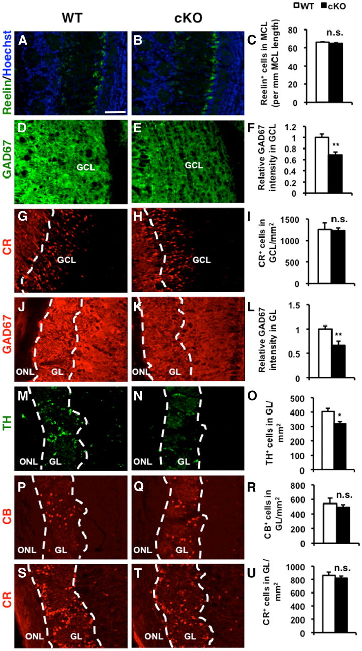
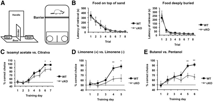
ERK5 MAP kinase is highly expressed in the developing nervous system and has been implicated in promoting the survival of immature neurons in culture. However, its role in the development and function of the mammalian nervous system has not been established in vivo. Here, we report that conditional deletion of the erk5 gene in mouse neural stem cells during development reduces the number of GABAergic interneurons in the main olfactory bulb (OB). Our data suggest that this is due to a decrease in proliferation and an increase in apoptosis in the subventricular zone and rostral migratory stream of ERK5 mutant mice. Interestingly, ERK5 mutant mice have smaller OB and are impaired in odor discrimination between structurally similar odorants. We conclude that ERK5 is a novel signaling pathway regulating developmental OB neurogenesis and olfactory behavior.


Figures

Figure 1.
Figure 1.
ERK5 protein expression in WT and cKO littermates. Coronal sections were prepared from E13.5, E16.5, P0, P28, and 6 month-old (6Mo) brains and immunostained with an affinity-purified anti-ERK5 antibody. A–G, ERK5 protein expression in WT mouse brains. H–N, ERK5 protein expression in cKO mouse brains. Insets, A–N, Hoechst-stained images to orient the location of the images. A′–N′, High-magnification images of the corresponding boxed area (A–N). D, E, K, L, Arrows point to the SVZ. LGE, Lateral ganglionic eminence; LV, lateral ventricle; GL, glomerular layer; GCL, granule cell layer. Scale bars: (in A) B–N′, 200 μm; (in A′) B′–N′, 40 μm.
Figure 2.
Figure 2.
ERK5 cKO mice have smaller OB. A, ERK5 deletion does not affect the overall lamina structure of the OB at P28. Images are Nissl-stained coronal sections. Scale bar, 200 μm. B, ERK5 deletion results in a smaller OB at P28. Data are unbiased stereological measurements of the OB volume from ERK5 cKO, ERK5 WT, or heterozygous (het) littermates. C, Stereological measurement of the volumes for each layer of the OB at P28. D, ERK5 cKO mice still have smaller OB at 6 months of age. ONL, Olfactory nerve layer; GL, glomerular layer; EPL, external plexiform layer; MCL, mitral cell layer; IPL, internal plexiform layer; GCL, granule cell layer. n = 5 per genotype. n.s., not significant; *p < 0.05; **p < 0.01.
Figure 3.
Figure 3.
ERK5 cKO mice have fewer neurons in the OB. A, Representative images of NeuN immunostaining in the granule cell layer (GCL) and glomerular layer (GL) of the OB from P28 ERK5 WT and cKO mice. B, C, Stereological quantification of NeuN+ cell density in GCL (B) and GL (C). Scale bar: (in A) A–C, 100 μm. *p = 0.05; p = 0.01.
Figure 4.
Figure 4.
ERK5 deletion reduces the number of GABAergic interneurons in the OB. Coronal sections of the OB from P28 ERK5 WT and cKO mice were analyzed. A–C, ERK5 deletion does not change the cell density of mitral cells, identified by anti-Reelin immunostaining. Hoechst staining (blue) was used to identify all nuclei and overall structure. D–F, ERK5 deletion significantly reduces immunoreactivity for GAD67, a marker for GABAergic neurons, in the granule cell layer (GCL). G–I, ERK5 deletion does not alter the cell density of Calretinin+ (CR+) cells in GCL. J–L, ERK5 deletion also reduces immunoreactivity for GAD67 in the glomerular layer (GL). M–O, ERK5 cKO mice have fewer TH+ dopaminergic neurons, a subpopulation of interneurons in the GL. P–U, ERK5 deletion does not affect the two interneuron subpopulations in the GL that express calcium binding proteins, Calbindin (CB; P–R) or Calretinin (CR; S--U). Scale bar: (in A) A–U, 100 μm. ONL, Olfactory nerve layer. n.s., not significant; *p < 0.05; **p < 0.01.
Figure 5.
Figure 5.
ERK5 deletion attenuates proliferation of progenitor cells in the anterior SVZ and RMS. ERK5 WT and cKO pups at the age of P0, P7, and P14 were dosed with BrdU and killed 2 h later. A, B, Montaged images of BrdU immunostaining of parasagittal sections of ERK5 WT (A) and cKO (B) brains at P0. Scale bar: (in A) A, B 500 μm. C, D, High-magnification images of yellow-boxed areas in A and B, respectively. Scale bar: (in C) C, D, 100 μm. E, Stereological quantification of total number of BrdU+ cells in the areas of anterior SVZ (SVZa)-RMS. F, Montaged images of Hoechst-stained parasagittal sections of P0 brains to visualize the area of anterior SVZa-RMS. The ERK5 cKO brain displays an enlarged lateral ventricle and smaller SVZa-RMS. Scale bar, 500 μm. G, Stereological quantification of the volume of SVZa-RMS of ERK5 WT and cKO mice. ERK5 deletion reduces the size of SVZa-RMS, the neurogenic region where OB interneurons originate. H, Representative images of Hoechst-stained coronal sections of P0 and P28 brains to visualize the size of the lateral ventricles. Scale bar, 200 μm. I, Stereological quantification of the volume of lateral ventricles of ERK5 WT and cKO mice at P0 and P28. The ERK5 cKO brain displays an enlarged lateral ventricle at P0 but not at P28. LV, Left ventricle. n.s., not significant; *p < 0.05; **p < 0.01.
Figure 6.
Figure 6.
ERK5 deletion does not change the stem cell pool in the SVZ at P28. A, Montaged images of Sox2 immunostained coronal sections of ERK5 WT and cKO brains at P28. Scale bar, 200 μm. Insets, High-power images of the corresponding boxed area. Scale bar: (in inset) 40 μm. B, Quantification of the accumulative Sox2 staining intensity in the entire SVZ of WT and cKO brains. n.s., Not significant.
Figure 7.
Figure 7.
ERK5 deletion does not affect neuroblast migration to the OB. Parasagittal sections of P14 ERK5 WT and cKO brains were subjected to PSA-NCAM immunostaining. A, Representative images of PSA-NCAM staining on three locations—SVZa, RMS, and the core of the OB corresponding to the three boxed areas in the montaged Hoechst-stained image—along the SVZa-RMS-OB path. Scale bar, 100 μm. B, Quantification of the PSA-NCAM staining intensity. n.s., Not significant.
Figure 8.
Figure 8.
ERK5 deletion increases apoptosis in both the anterior SVZ-RMS and the OB. Parasagittal sections of WT and ERK5 cKO brains at P0 and P7 were subjected to TUNEL and/or active caspase3 immunostaining to analyze the apoptosis in SVZa-RMS. Coronal sections of P28 brains were subjected to TUNEL to examine the apoptosis in the granule cell layer (GCL) and glomerular layer (GL) of the OB. A, Representative images of TUNEL (green) in the anterior SVZ of P0 brain. Hoechst staining (blue) was used to identify all nuclei and overall structure. Scale bar,100 μm. B, Quantification of total number of TUNEL+ cells per SVZa-RMS. C, Representative images of active caspase3 immunostaining (green) in the anterior SVZ of P0 brain. Scale bar, 100 μm. D, Quantification of total number of active caspase3+ cells per SVZa-RMS. E, Representative images of TUNEL (green) in the GCL of the OB at P28. Scale bar, 50 μm. F, Quantification of total number of TUNEL+ cells in GCL and GL per OB. *p < 0.05; **p < 0.01.
Figure 9.
Figure 9.
ERK5 deletion does not affect the development of the main olfactory epithelium (MOE), the EOG response of the MOE to odorants, or the innervation of the OB. A, ERK5 deletion does not change the thickness of the MOE, measured after Nissl staining. B, ERK5 deletion does not alter the immunoreactive profile of AC3 in the cilia layer of the MOE. Scale bar, 50 μm. C, Odorant-stimulated EOG responses were similar in the MOE prepared from ERK5 WT and cKO mice. The MOE tissue was air-puffed with H2O, citralva (1 mm), ethyl vanillin (100 μm), limonene (−) (10 mm), and IAA (1 mm). The membrane potential was recorded by EOG. Arrows point to the start of odorant administration. D, Summary of the mean EOG amplitudes in response to odorants. Citra, citralva; Vani, ethyl vanillin; Lim, limonene (−); IAA, isoamyl acetate. E, F, ERK5 deletion does not change the immunostaining pattern or intensity for olfactory marker protein (OMP) in the OB of P28 mice. OMP is a marker of mature olfactory sensory neurons that project to the olfactory nerve layer (ONL) and glomerular layer (GL). Scale bar: (in E) E, F, 200 μm. G, H, Representative images of OMP immunostaining of the OB from P0 pups. The pattern of innervation of the OB by olfactory sensory neurons is similar between ERK5 WT and cKO pups at P0. Scale bar: (in G) G, H, 400 μm. I, J, High-magnification images of the boxed areas in G and H, respectively, showing the development of glomerulus-like structures (arrows) in the OB from both ERK5 WT and cKO pups. Scale bar: (in I) I, J, 100 μm. n.s., Not significant.
Figure 10.
Figure 10.
ERK5 cKO mice are deficient in the discrimination of structurally similar odors under a cotton tip presentation-based task. A, ERK5 cKO mice are normal in the standard habituation/dishabituation olfaction test. Naive, adult mice were pretrained with four presentations of mineral oil-soaked cotton swabs, then exposed to three structurally different odorants—octanol, benzaldehyde, and ethyl vanillin—with 4 trials per odorant. Both WT and ERK5 cKO mice were able to detect and discriminate between the distinct odorants. B, ERK5 cKO mice cannot distinguish between the structurally similar limonene (+) and limonene (−). Animals were exposed to four presentations of limonene (+) followed by one presentation to limonene (−). WT, but not ERK5 cKO mice, sniffed limonene (−) significantly more than the fourth presentation of limonene (+), suggesting their ability to discriminate between the two similar odorants. C, Similarly, unlike their WT littermates, ERK5 cKO mice cannot discriminate between butanol and pentanol. D, Neither ERK5 WT nor cKO mice show any preference toward a particular odorant between each pair of the structurally similar ones. Animals were presented simultaneously with two cotton tips laced with a pair of the structurally similar odorants, limonene (+) versus (−), or butanol versus pentanol. Lim, limonene; Buta, butanol; Penta, pentanol. n ≥ 10 for WT and n = 9 for cKO. n.s., Not significant; **p < 0.01.
Figure 11.
Figure 11.
ERK5 cKO mice are deficient in the discrimination of structurally similar odors under a sand digging-based task. A, Schematic diagram of the behavior apparatus and set up for the assay. The animal was confined to one end of his home cage by a barrier while the apparatus was lowered to the other end of the cage to deliver the sand-filled dishes and food. The barrier was then removed to allow the animal to investigate the sand dishes and retrieve the food. B, Both ERK5 cKO mice and WT littermate controls learned to retrieve the food reward that is placed on top of the sand or deeply buried in the sand during pretraining sessions. C, Mice were trained to associate an odor cue with the food reward and discriminate two structurally unrelated odors, IAA and citralva. The food reward was buried deeply under the sand scented with IAA. D, ERK5 cKO mice are deficient in distinguishing between the structurally similar limonene (+) versus limonene (−). The food reward was buried deeply under the sand scented with limonene (−). E, Unlike their WT littermates, ERK5 cKO mice cannot discriminate between butanol and pentanol. The food reward was buried deeply under the sand scented with pentanol. n = 10 and 9 for WT and cKO, respectively. *p = 0.05; **p = 0.01.

References

    1. Abraham NM, Egger V, Shimshek DR, Renden R, Fukunaga I, Sprengel R, Seeburg PH, Klugmann M, Margrie TW, Schaefer AT, Kuner T. Synaptic inhibition in the olfactory bulb accelerates odor discrimination in mice. Neuron. 2010;65:399–411. - PMC - PubMed
    1. Adam Y, Mizrahi A. Circuit formation and maintenance–perspectives from the mammalian olfactory bulb. Curr Opin Neurobiol. 2010;20:134–140. - PubMed
    1. Allen ZJ, 2nd, Waclaw RR, Colbert MC, Campbell K. Molecular identity of olfactory bulb interneurons: transcriptional codes of periglomerular neuron subtypes. J Mol Histol. 2007;38:517–525. - PubMed
    1. Alvarez-Buylla A, Garcia-Verdugo JM. Neurogenesis in adult subventricular zone. J Neurosci. 2002;22:629–634. - PMC - PubMed
    1. Batista-Brito R, Close J, Machold R, Fishell G. The distinct temporal origins of olfactory bulb interneuron subtypes. J Neurosci. 2008;28:3966–3975. - PMC - PubMed

Publication types

MeSH terms

LinkOut - more resources