. 2012 Mar 21;32(12):4284-96.
doi: 10.1523/JNEUROSCI.6332-11.2012.

P2X2 and P2X5 subunits define a new heteromeric receptor with P2X7-like properties

Affiliations

P2X2 and P2X5 subunits define a new heteromeric receptor with P2X7-like properties

Vincent Compan et al. J Neurosci. .

Abstract

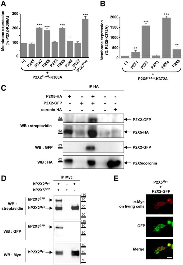
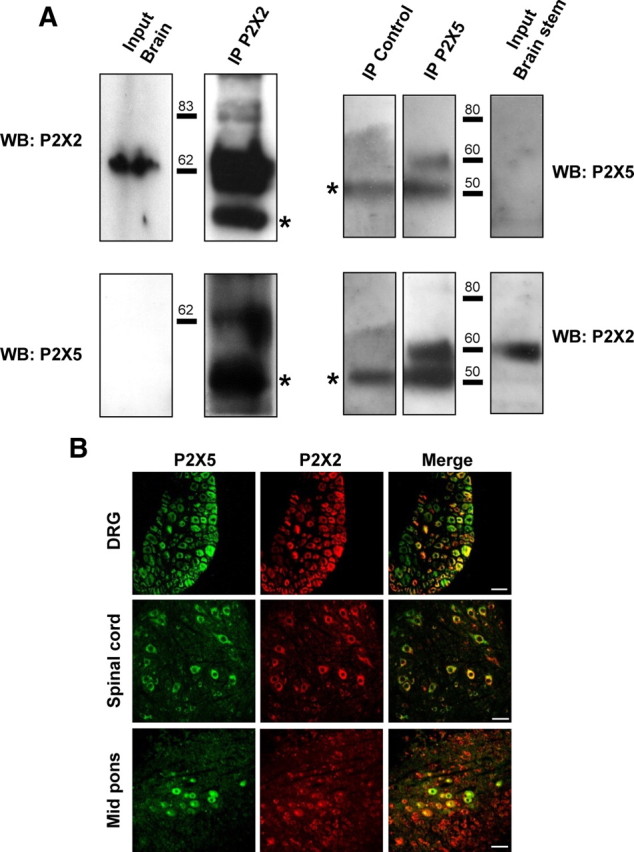
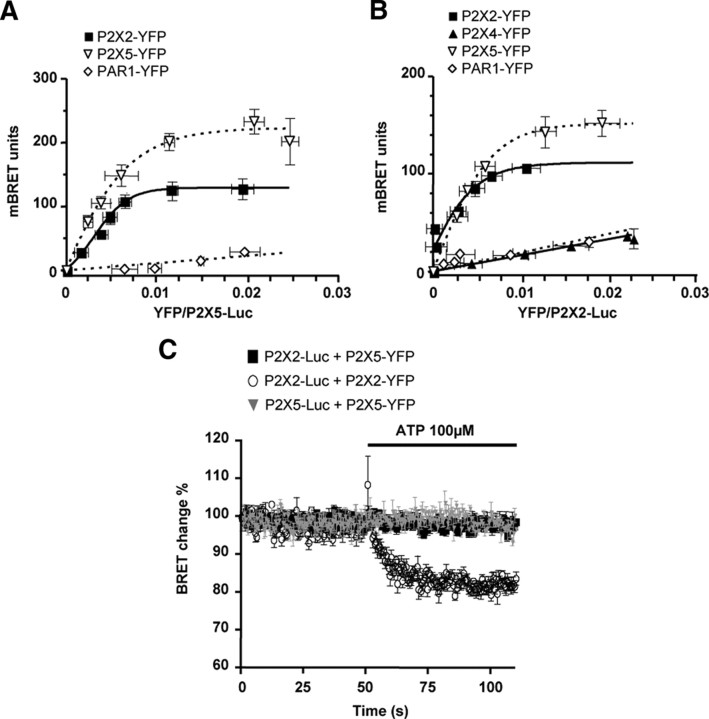
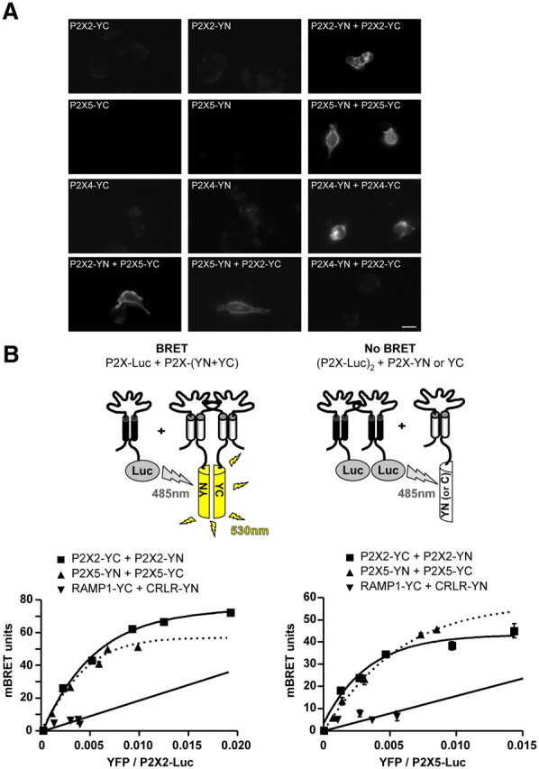
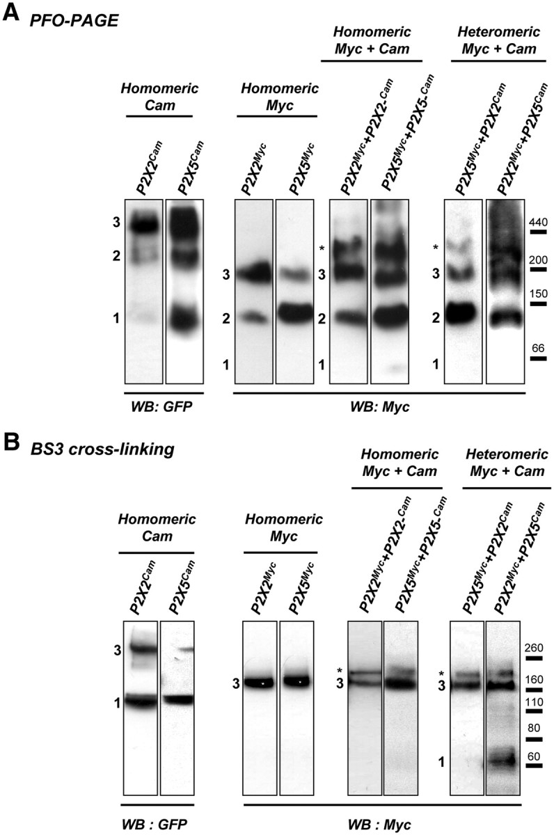
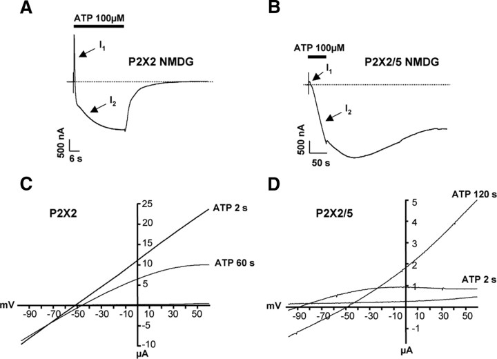
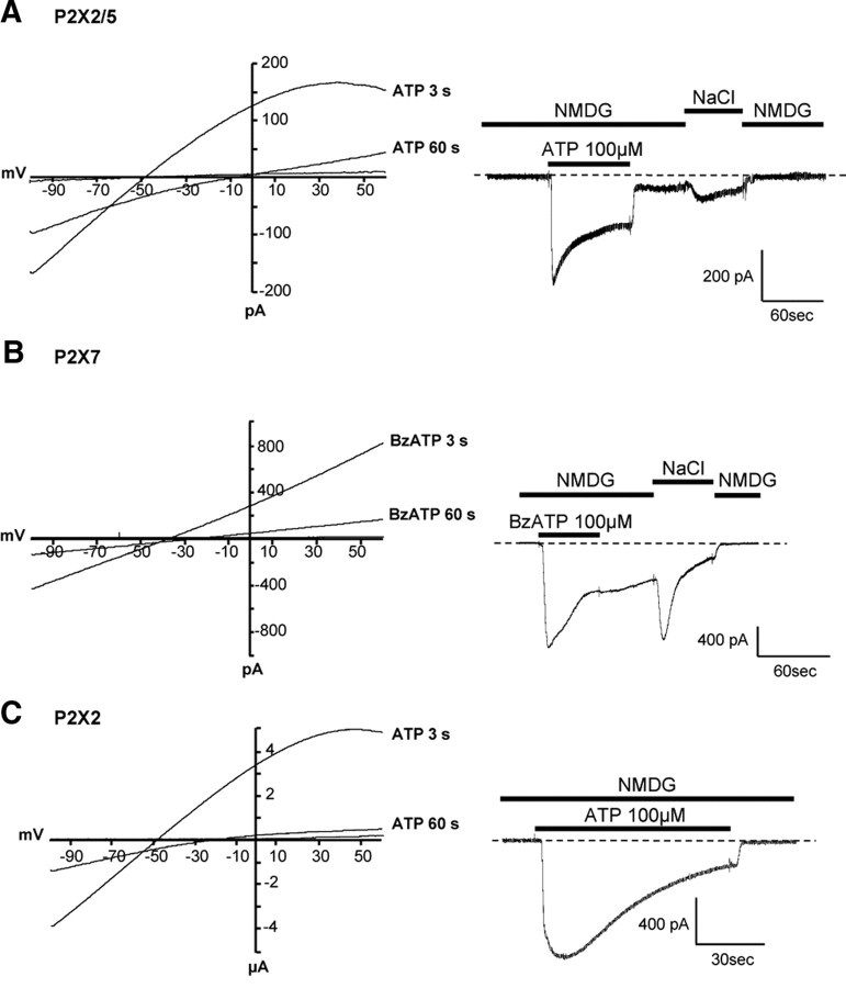
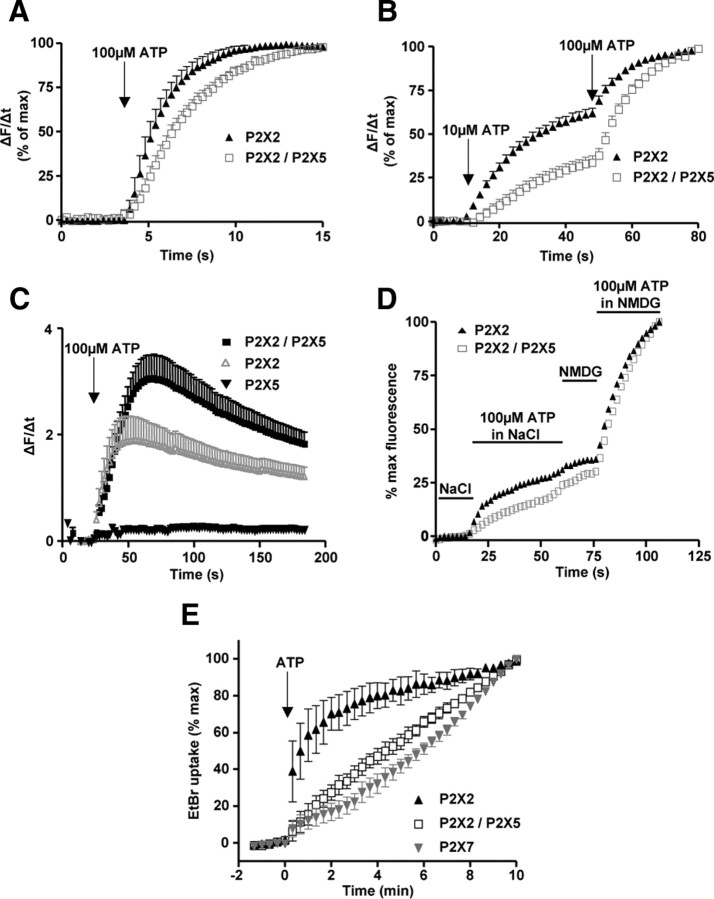
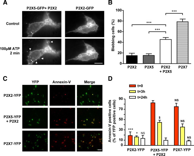
Ligand-gated ion channels are prototypic oligomeric membrane proteins whose stoichiometry determines their functional properties and subcellular localization. Deciphering the quaternary structure of such protein complexes is an arduous task and usually requires the combination of multiple approaches. ATP-gated P2X receptors are formed by the association of three subunits, but the quaternary arrangement of the seven P2X subunits at the plasma membrane remains poorly characterized. By combining bioluminescence resonance energy transfer, bifunctional fluorescence complementation and protein biochemistry, we developed an experimental approach that allows precise determination of rat P2X receptor quaternary assembly. We found that P2X5 subunits associate with P2X1, P2X2, and P2X4 subunits. We demonstrate that P2X5 and P2X2 subunits interact to form as yet uncharacterized heteromeric receptors with alternate stoichiometries, both present at the plasma membrane. P2X2/5 receptors display functional properties such as pore dilatation, membrane blebbing, and phosphatidylserine exposure that were previously thought to be characteristic hallmarks of the P2X7 receptor. In mouse, P2X2 and P2X5 subunits colocalize and physically interact in specific neuronal populations suggesting that other P2X receptors might contribute to cellular responses typically attributed to P2X7 receptor.


Figures

Figure 1.
Figure 1.
P2X5 subunits interact with P2X2 subunits at the plasma membrane. A, Rescue of cell surface expression of the P2X2–K366A trafficking deficient mutant by other subunits. P2X2–K366A carrying a Flag tag in the extracellular loop was expressed alone or in combination with each of the seven wild-type P2X subunits. Membrane expression was measured using a chemiluminescent assay. Cell surface expression of P2X2–K366A is rescued upon coexpression with P2X2, P2X3, P2X4, and P2X5 subunits. B, Membrane detection of the P2X5–K372A mutant carrying an extracellular Flag tag is increased when coexpressed with P2X1, P2X2, and P2X4 and P2X5 subunits. Note that because the surface expression of P2X5–K372 mutant is very low compared withP2X2–K366A, the changes appear much higher. Results are shown as mean ± SEM of at least three independent experiments. **p < 0.01, ***p < 0.005, Student's t test. C, P2X5 subunits interact with P2X2 at the plasma membrane. P2X5 carrying an extracellular HA tag was expressed alone or in combination with P2X2-GFP. Immunoprecipitation was performed after labeling living cells with sulfo-NHS-LC-biotin. Top, Biotinylated protein fraction after HA immunoprecipitation detected with streptavidin-HRP. Middle and bottom, Detection of coimmunoprecipitated GFP and HA tagged-proteins, respectively. Coronin was used as a control for cell permeabilization. D, Human P2X2 and P2X5 subunits interact at the plasma membrane. Experiments were carried as described above, except that human P2X2-Myc and human P2X5-GFP cDNAs were used. E, Immunodetection of P2X5 in living cells. HEK cells were transfected with P2X5Myc subunits carrying an extracellular Myc tag and P2X2-GFP. Immunostaining of P2X5Myc subunits (red) was performed by incubation of Myc antibodies on living cells; P2X2 subunits (green) were revealed through GFP fluorescence. Scale bar, 10 μm.
Figure 2.
Figure 2.
P2X5 and P2X2 subunits interact in neuronal tissues. A, Physical interactions of P2X2 and P2X5 subunits in brain tissues. P2X2 or P2X5 subunits were immunoprecipitated from mouse brain membrane and different subunits detected with specific antibodies. Left panel represents immunoprecipitation from total brain membrane proteins, right panel from brainstem. Note that P2X5 is not detectable from the total protein extract. Blots are representative of three independent experiments. Asterisks indicate the antibody heavy chains. B, Coimmunolocalization of P2X2 and P2X5 subunits in peripheral and central neurons. Representative images of immunohistochemistry performed on slices from DRG, spinal cord, or mid pons. P2X2 and P2X5 are stained in red and green, respectively. Note that in each structure only a subset of neurons coexpresses both subunits. Scale bar, 40 μm.
Figure 3.
Figure 3.
P2X5 subunits are in close spatial proximity to P2X2 subunits. A, BRET titration curves using P2X5-Luc as the energy donor. HEK cells were cotransfected with a constant amount of P2X5-Luc and with an increasing amount of YFP fusions. BRET signals are plotted against the relative amounts of each tagged subunit. Specific, saturating, BRET signals are observed between P2X5 and P2X2, or P2X5 subunits, while low linear BRET is obtained with PAR-1. B, BRET titration curves using P2X2-Luc as the energy donor, with experiments performed as described above. Specific BRET signals are observed between P2X2 and P2X2 or P2X5 subunits, but not P2X4 and PAR-1. Data are expressed as mean ± SEM of at least N = 3 experiments. C, Cytosolic domains of P2X2/5 and P2X2 receptors undergo differential conformational changes after ATP stimulation. Dynamic BRET signals were recorded after ATP stimulation (100 μm) in HEK cells transfected with P2X2, and P2X5 subunits carrying either a luciferase (Luc) or YFP tag on their C terminus tail, alone or in combination. ATP stimulation induced a diminution of BRET signal between P2X2 subunits, which is not observed for P2X2/5 and P2X5 receptors. Representative experiment reproduced three times. Data are mean ± SEM of triplicate.
Figure 4.
Figure 4.
Stoichiometry of P2X2 and P2X5 interactions as assessed by BiFC and BRET/BiFC. A, Bimolecular fluorescence complementation between P2X subunits. P2X subunits fused to either the amino or carboxyl half of YFP (YN or YC) were transfected alone or in combination in HEK cells. Recomplemented fluorescence was observed by microscopy. BiFC was observed between homomeric P2X2, P2X4, and P2X5 subunits, as well as between heteromeric P2X2 and P2X5 subunits, but not between P2X2 and P2X4 subunits. Scale bar, 10 μm. B, Combination of BRET and BiFC reveals the stoichiometry of the heteromeric P2X2/P2X5 assembly. Top, Diagram illustrating the approach. BRET is only observed between one P2X2-Luc subunit and any other combinations of two P2X-hemi-YFP subunits. BRET titration curves between P2X2-Luc (left) or P2X5-Luc (right) cotransfected with P2X2, P2X5, RAMP1, and CRL fused to hemi-YFP. Specific BRET signals were observed between P2X2 and P2X5 subunits for each experimental condition, demonstrating the existence of heteromeric receptors with two different stoichiometries. Data are expressed as mean ± SEM of at least N = 3 experiments.
Figure 5.
Figure 5.
Biochemical analysis of P2X2/5 subunit assembly. A, Analysis of the P2X2/5 subunit complex in native conditions using PFO-PAGE. HEK cells were transfected with P2X subunits with either a Myc tag or the Cam protein fused to their C terminus. Homomeric P2X2 and P2X5 receptors fused to Myc or Cam are resolved as monomers, dimers, and trimers (indicated by numbers). When Myc and Cam fused P2X subunits are coexpressed, an additional high molecular weight form of the complex (indicated by an asterisk) is detected, which corresponds to a stoichiometry of two Myc subunits for one Cam subunit. Molecular weight markers apoferritin (440 kDa), β-amylase (200 kDa), alcohol dehydrogenase (150 kDa), and bovine serum albumin (66 kDa) were detected by Ponceau red staining before membrane blocking. B, P2X2/5 receptors are present at the plasma membrane with two different stoichiometries. Stoichiometry of P2X receptors at the plasma membrane was analyzed after living cell surface protein cross-linking with the membrane impermeable cross-linker BS3. Cross-linked P2X subunits were analyzed by SDS/PAGE. Homomeric Myc-tagged receptors are resolved as a single band corresponding to the molecular weight of a trimer, while Cam-tagged subunits migrate as monomers and trimers. When Myc- and Cam-fused P2X subunits are coexpressed, an additional high molecular weight form of the cross-linked receptor (indicated by an asterisk) is detected, corresponding to a stoichiometry of one P2X-Cam subunit and two P2X-Myc subunits. All data are representative of N > 3 independent experiments.
Figure 6.
Figure 6.
P2X2/5 receptor stimulation evokes sustained NMDG conductance in Xenopus oocytes. Permeability dynamics of P2X2 (A) and P2X2/5 (B). Representative currents evoked by ATP (100 μm) in Xenopus oocytes expressing P2X2 or P2X2/5 receptors recorded in NMDG external solution at a holding potential of −60 mV. I1 and I2 correspond to channels permeant to small cations or NMDG+, respectively. Note the faster I1 to I2 transition for P2X2 receptors compared with P2X2/5, and the lack of reversibility of the inward P2X2/5 conductance. C, D, Shift in the reversal potential of ATP-evoked currents in NMDG extracellular solution. Currents were measured during repeated voltage ramps protocol in the presence of 100 μm ATP. Two seconds after ATP application, reversal potential of P2X2 current has already changed compared with that of P2X2/5. Note the difference in current rectification between the two types of channels.
Figure 7.
Figure 7.
P2X2/5 and P2X7 NMDG conductances display similar properties in HEK cells. Left, Shift in the reversal potential of ATP-evoked currents in NMDG extracellular solution in HEK cells expressing P2X2/5 (A), P2X7 (B), or P2X2 (C). Currents elicited by 100 μm ATP were recorded during 100 ms voltage ramps from −100 to +80 mV, applied from a holding potential of −60 mV. Right, Representative currents recorded in NMDG external solution evoked by 100 μm ATP (or 100 μm BzATP for P2X7) in HEK cells expressing P2X2/5 (A), P2X7 (B) or P2X2 (C). Holding potential −60 mV. P2X2/5 and P2X7-expressing cells show a NMDG conductance that persists after agonist washout and can be fully blocked by extracellular NaCl. Note that in P2X2-expressing cells, the NMDG conductance readily reverses upon agonist washout.
Figure 8.
Figure 8.
Activation of P2X2/5 receptors induces dye uptake. A, B, ATP-evoked Yo-Pro-1 uptake in HEK cells expressing P2X2 or P2X2/5 receptors. Video-microscopy experiments were performed in NMDG extracellular solution. Individual cell fluorescence signals were differentiated and normalized to the maximal response. Data are mean ± SEM of n > 10 cells, N = 3–4 experiments. Note that the kinetics of Yo-Pro-1 uptake are slower for P2X2/5 receptor compared with P2X2. C, P2X2/5 displays greater ATP-evoked Yo-Pro-1 uptake compared with P2X2. Experiments were performed as above, except that results were not normalized. Data are mean ± SEM of n > 10 cells, N = 4 experiments. D, ATP-evoked Yo-Pro1 uptake occurs in physiological solution. Cells were stimulated as above, first in physiological solution (NaCl-based solution) and then in NMDG-based solution. In both conditions, ATP evoked Yo-Pro-1 uptake albeit with higher potency in NMDG solution. Data are mean of n > 10 cells, N = 3 experiments. SEM was omitted for clarity. E, Comparison of ethidium bromide uptake evoked by P2X2, P2X2/5, or P2X7 receptors stimulation. Cells were stimulated with 100 μm ATP (5 mm for P2X7) in NaCl-based solution. Results are mean ± SEM, n > 30 cells, N = 4 experiments. Note that P2X2/5- and P2X7-evoked EtBr uptakes do not saturate.
Figure 9.
Figure 9.
Activation of P2X2/5 receptors mediates membrane blebbing and pseudo-apoptosis. A, B, P2X2/5 activation triggers membrane blebbing. A, Representative fluorescence images of HEK cells expressing P2X2-GFP or P2X2 + P2X5-GFP 2 min after 100 μm ATP stimulation. Arrows indicate ATP induced blebs. Scale bar, 10 μm. B, Quantitative analysis of the percentage of blebbing cells expressing P2X2-GFP, P2X5-GFP, P2X2/5-GFP, or P2X7-GFP after stimulation with 100 μm ATP (2 mm for P2X7) for 2 min. Results were normalized to the number of GFP-positive cells. Data are mean ± SEM of N = 3 independent experiments. ***p < 0.005, one-way ANOVA, followed by Bonferroni's multiple-comparison test. C, D, P2X2/5 receptor activation induces transient annexin-V exposure. C, HEK cells transfected with P2X2-YFP, P2X2+P2X5-YFP, or P2X7-YFP were stimulated with 100 μm ATP (500 μm for P2X7-YFP) and annexin-V staining was performed 5 min after the end of the stimulation. Scale bar, 50 μm. D, Analysis of annexin-V staining at 0, 3, or 24 h after ATP stimulation. Experiment was performed as in C, except that annexin-V staining was performed at the time indicated after ATP stimulation. In all panels, results were normalized to the number of YFP-positive cells. Data are mean ± SEM, n > 30 cells, N = 3 experiments. *p < 0.05, ***p < 0.005; N.S., not significant by comparison with P2X2/5 transfected cells at the same time point. $p < 0.05, comparison between P2X2/5-expressing cells at 0 and 3 h. One-way ANOVA, followed by Bonferroni's multiple-comparison test.

References

    1. Anderson CM, Nedergaard M. Emerging challenges of assigning P2X7 receptor function and immunoreactivity in neurons. Trends Neurosci. 2006;29:257–262. - PubMed
    1. Antonio LS, Stewart AP, Xu XJ, Varanda WA, Murrell-Lagnado RD, Edwardson JM. P2X4 receptors interact with both P2X2 and P2X7 receptors in the form of homotrimers. Br J Pharmacol. 2011;163:1069–1077. - PMC - PubMed
    1. Aschrafi A, Sadtler S, Niculescu C, Rettinger J, Schmalzing G. Trimeric achitecture of homomeric P2X2 and heteromeric P2X1+2 receptor subtypes. J Mol Biol. 2004;342:333–343. - PubMed
    1. Ayoub MA, Maurel D, Binet V, Fink M, Prézeau L, Ansanay H, Pin JP. Real-time analysis of agonist-induced activation of protease-activated receptor 1/Galphai1 protein complex measured by bioluminescence resonance energy transfer in living cells. Mol Pharmacol. 2007;71:1329–1340. - PubMed
    1. Barrera NP, Henderson RM, Murrell-Lagnado RD, Edwardson JM. The stoichiometry of P2X2/6 receptor heteromers depends on relative subunit expression levels. Biophys J. 2007;93:505–512. - PMC - PubMed

Publication types

MeSH terms