. 2012 Jul;93(Pt 7):1563-1572.
doi: 10.1099/vir.0.039354-0. Epub 2012 Mar 21.

Type-specific interaction between human papillomavirus type 58 E2 protein and E7 protein inhibits E7-mediated oncogenicity

Affiliations

Type-specific interaction between human papillomavirus type 58 E2 protein and E7 protein inhibits E7-mediated oncogenicity

Xin Wang et al. J Gen Virol. 2012 Jul.

Abstract

Human papillomavirus type 58 (HPV-58) is a very common HPV type in eastern Asia. Little is known about its biology and tumorigenesis. In this study, HPV-58 E2 protein (58E2) was found to interact with E7 protein (58E7), and the hinge domain of 58E2 was shown to be responsible for binding to the 58E7 protein. Interestingly, the E2-E7 interaction appears to be HPV type-specific, as we found that the HPV-16 E2 could not bind to the 58E7 protein, and neither did 58E2 interact with HPV-16 E7. The biological consequence(s) of the E2-E7 interaction in HPV-58, especially in viral tumorigenesis, was investigated. Results showed that, through interacting with 58E7, 58E2 prevented E7-induced retinoblastoma protein (pRb) degradation and prolonged the half-life of pRb in cells. Additionally, 58E2 abrogated 58E7-induced cell proliferation. These observations collectively suggest that direct interaction with 58E7 is another mechanism for 58E2 to inhibit 58E7-associated carcinogenesis in addition to regulating expression of the 58E7 gene.


Figures

Fig. 1.
Fig. 1.
Interaction between HPV 58E2 and 58E7 proteins. (a) The 58E2 fragments [NT (aa 1–201) and HDCT2 (aa 225–358)] fused with GST and 58E7 protein fused with 6×His tag were used in a GST pull-down assay as described in Methods. The pull-down proteins were subjected to Western blot analysis (WB) with an anti-His antibody (lower panel). Input purified proteins (2 %) were analysed by WB, employing anti-GST (upper panel) and anti-His (middle panel) antibodies for the detection of GST–58E2 mutants and His–58E7, respectively. (b) 293T cell monolayers were co-transfected with Flag-tagged E7-expressing plasmids (pCMV-3Tag-1A-16E7/58E7) and Myc-tagged E2-expressing plasmids (pCMV-3Tag-2A-16E2/58E2). Twenty-four hours post-transfection, whole-cell extracts (WCE) were prepared from the transfected cells, and 4 % of the WCE was used to detect the expression of Myc–E2 and Flag–E7 proteins by WB, employing anti-Myc (upper panel) and anti-Flag (middle panel) antibodies, respectively. The remaining cell lysates were immunoprecipitated (IP) with a mouse-anti-Flag M2 affinity gel and the immunoprecipitates were resolved by SDS-PAGE and subjected to WB analysis with a rabbit anti-Myc antibody to detect the presence of E2 in the pellets (lower panel).
Fig. 2.
Fig. 2.
Mapping the domains of 58E2 binding to 58E7. (a) Schematic diagram of the 58E2 truncations used in this study. (b) 293T cells were co-transfected with a Flag-tagged 58E7-expressing plasmid and the expression vectors for the Myc-tagged 58E2 truncation mutants as indicated. Twenty-four hours post-transfection, whole-cell extracts (WCE) were prepared from the transfected cells, and the expression levels of Flag–58E7 and the Myc–58E2 mutants were analysed by Western blotting (WB). The cell lysates were subjected to immunoprecipitation (IP) with a mouse-anti-Flag M2 affinity gel followed by WB analysis with a rabbit-anti-Myc antibody to detect the presence of 58E2 mutants in the pellets (lower panel). Molecular mass standards (in kDa) are indicated on the right.
Fig. 3.
Fig. 3.
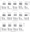
58E2 protects pRb from 58E7-induced degradation. HaCat cells were transiently co-transfected with the expression vectors of HPV E7 and HPV E2 proteins or 58E2 truncation mutants as indicated. 1A and 2A represent the empty vector pCMV-3tag-1A and pCMV-3tag-2A, respectively. Twenty-four hours post-transfection, protein biosynthesis was blocked by addition of 100 µg CHX ml−1 and cells were harvested and lysed at the indicated time points (0, 3, 6 h) after adding CHX. The equal amount of cell lysates protein was subjected to SDS-PAGE. The steady-state levels of pRb were determined by Western blot analysis (top panels; results shown are one of three independent experiments), and β-actin served as a loading control (lower panels). The densities of the pRb bands were normalized by comparison with the respective β-actin bands, and then the relative pRb values were expressed as a percentage where the value of 0 h CHX treatment in the same group corresponded to 100 %. Results shown are means±sd of three experiments. *P<0.05 (Student's t-test), pRb levels in CHX-treated cells for 0 h versus 6 h.
Fig. 4.
Fig. 4.
58E2 does not compete with pRb for binding to 58E7. HaCat cells were co-transfected with 58E2 (or the NT fragment) and 58E7 (or its mutant 58E7Δ22–26) expression vectors as indicated. Twenty-four hours post-transfection, whole-cell extracts (WCE) were prepared and the input proteins and cellular pRb were analysed by Western blotting (WB). The cell lysates were subjected to immunoprecipitation (IP) with a mouse-anti-Flag antibody that recognizes Flag-tagged E7 proteins. Myc–58E2 or pRb was detected in the immunoprecipitates with rabbit anti-Myc-tag or rabbit anti-pRb antibody, respectively. The empty vector (E) pCMV-3tag-1A and 58E2 co-transfection set served as a blank control.
Fig. 5.
Fig. 5.
Inhibitory effects of 58E2 on 58E7-induced cell colony formation. The colony-formation assay was performed as described in Methods. Briefly, HaCat cells were co-transfected as indicated. Cells co-transfected with empty vectors ‘1A+2A’ served as a blank control, while the 16E7-transfected and ‘16E7+16E2’ co-transfected cells served as a pair of positive controls. At 48 h post-transfection, 1×10⁴ cells of each set were resuspended and cultured in six-well plates, each well making two duplicates. Cells were cultured in Dulbecco’s modified Eagle’s medium (DMEM) containing 10 % FBS and 500 µg G418 ml−1. (a) After 14 days, the colonies were fixed with 3.7 % paraformaldehyde and stained by crystal violet. (b) The numbers in the graph are the mean±sd numbers of colonies from three independent experiments. *Values significantly different at P<0.05 (Student's t-test or ANOVA test).

Similar articles

Cited by

References

    1. Androphy E. J., Lowy D. R., Schiller J. T. (1987). Bovine papillomavirus E2 trans-activating gene product binds to specific sites in papillomavirus DNA. Nature 325, 70–73 10.1038/325070a0 - DOI - PubMed
    1. Blachon S., Bellanger S., Demeret C., Thierry F. (2005). Nucleo-cytoplasmic shuttling of high risk human papillomavirus E2 proteins induces apoptosis. J Biol Chem 280, 36088–36098 10.1074/jbc.M505138200 - DOI - PubMed
    1. Boyer S. N., Wazer D. E., Band V. (1996). E7 protein of human papilloma virus-16 induces degradation of retinoblastoma protein through the ubiquitin-proteasome pathway. Cancer Res 56, 4620–4624 - PubMed
    1. Breiding D. E., Sverdrup F., Grossel M. J., Moscufo N., Boonchai W., Androphy E. J. (1997). Functional interaction of a novel cellular protein with the papillomavirus E2 transactivation domain. Mol Cell Biol 17, 7208–7219 - PMC - PubMed
    1. Chan P. K., Cheung J. L., Cheung T. H., Lo K. W., Yim S. F., Siu S. S., Tang J. W. (2007). Profile of viral load, integration, and E2 gene disruption of HPV58 in normal cervix and cervical neoplasia. J Infect Dis 196, 868–875 10.1086/520884 - DOI - PubMed

Publication types

Substances

LinkOut - more resources