. 2012 Jun 15;125(Pt 12):3004-11.
doi: 10.1242/jcs.105114. Epub 2012 Mar 22.

VPS13 regulates membrane morphogenesis during sporulation in Saccharomyces cerevisiae

Affiliations

VPS13 regulates membrane morphogenesis during sporulation in Saccharomyces cerevisiae

Jae-Sook Park et al. J Cell Sci. .

Abstract

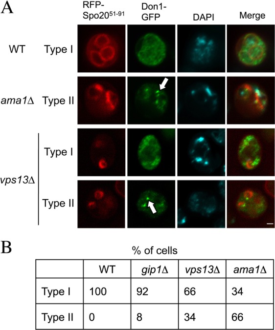
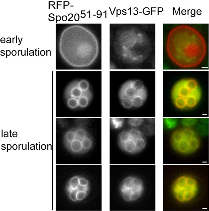
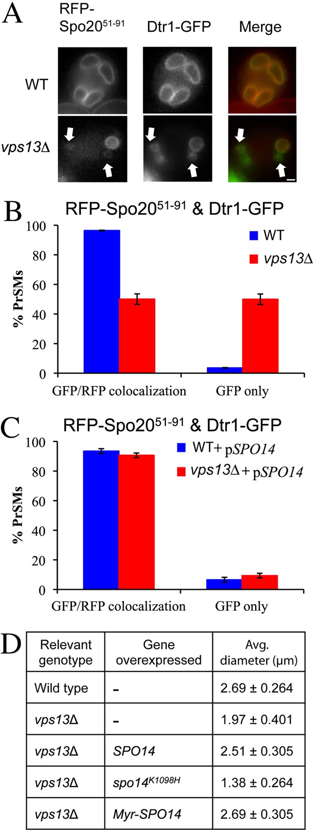
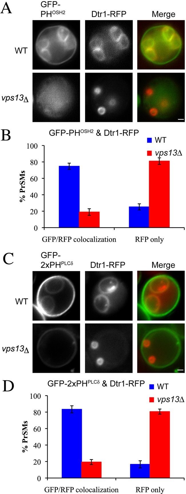
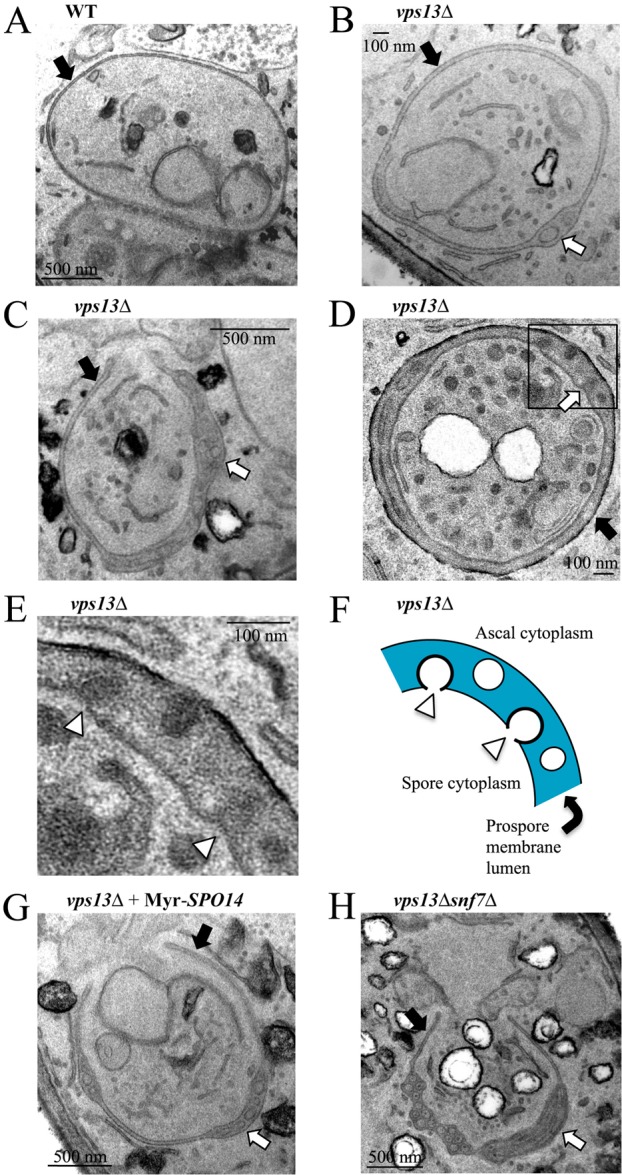
The hereditary disorders chorea acanthocytosis and Cohen syndrome are caused by mutations in different members of a family of genes that are orthologs of yeast VPS13. In vegetatively growing yeast, VPS13 is involved in the delivery of proteins to the vacuole. During sporulation, VPS13 is important for formation of the prospore membrane that encapsulates the daughter nuclei to give rise to spores. We report that VPS13 is required for multiple aspects of prospore membrane morphogenesis. VPS13 (1) promotes expansion of the prospore membrane through regulation of phosphatidylinositol phosphates, which in turn activate the phospholipase D, Spo14; (2) is required for a late step in cytokinesis that gives rise to spores; and (3) regulates a membrane-bending activity that generates intralumenal vesicles. These results demonstrate that Vps13 plays a broader role in membrane biology than previously known, which could have important implications for the functions of VPS13 orthologs in humans.


Figures

Fig. 1.
Fig. 1.
Don1 localization in vps13Δ cells. (A) Representative wild-type (AN120), gip1Δ (NY501), vps13Δ (HI29) and ama1Δ (ADY66) cells transformed with pRS426-RFP-Spo205191-DON1-GFP to visualize the prospore membranes and the LEP, respectively. Type I indicates that Don1–GFP was dispersed in the cytoplasm, whereas Type II indicates that Don1–GFP was concentrated on the prospore membrane. White arrows indicate LEPs. Scale bar: 1 µm. (B) Quantification of Don1–GFP distribution in the cells in A. More than 100 prospore membranes were examined for each strain.
Fig. 2.
Fig. 2.
Localization of Vps13–GFP in sporulating cells. A Vps13–GFP strain (JSP257) carrying pRS426-RFP-Spo205191 as a prospore membrane marker was sporulated and GFP and RFP fluorescence was monitored during prospore membrane growth. Scale bars: 1 µm.
Fig. 3.
Fig. 3.
Effects of vps13Δ on phosphatidic acid in the prospore membrane. (A) Wild-type (AN120) or vps13Δ (HI29) cells expressing both an integral membrane marker for the prospore membrane, DTR1-GFP, and the PA sensor RFP-Spo205191 were examined during sporulation. Arrows indicate Dtr1–GFP marked prospore membranes that lack RFP fluorescence. (B) Quantification of Dtr1–GFP and RFP–Spo2051–91 colocalization in the strains in A. At least 110 prospore membranes were scored in two independent experiments. (C) Wild-type (AN120) or vps13Δ (HI29) cells transformed with both pRS424-DTR1-GFP and pRS426-RFP-Spo205191-SPO14 were analyzed as in B. (D) The diameter of the prospore membrane was measured in post-meiotic wild-type (AN120) or vps13Δ (HI29) cells overexpressing the indicated genes. More than 100 prospore membranes were measured for each strain. The numbers given are the average diameter ± one standard deviation. Scale bar: 1 µm.
Fig. 4.
Fig. 4.
Effects of vps13Δ on prospore membrane PtdIns(4)P and PtdIns(4,5)P2 pools. (A) Wild-type (AN120) and vps13Δ (HI29) cells expressing the PtdIns(4)P sensor GFP–PHOSH2 and the prospore membrane marker Dtr1–RFP were examined during sporulation. (B) Quantification of GFP–PHOSH2 and Dtr1–RFP colocalization in A. (C) Wild-type (AN120) and vps13Δ (HI29) cells expressing the PtdIns(4,5)P2 sensor GFP–2XPHPLCδ and the prospore membrane marker Dtr1–RFP were examined during sporulation. (D) Quantification of GFP–2XPHPLCδ and Dtr1–RFP colocalization in C. More than 100 prospore membranes were observed for each strain. Scale bars: 1 µm.
Fig. 5.
Fig. 5.
A previously unknown prospore membrane phenotype in vps13Δ cells. TEM images of prospore membranes in (A) wild-type (AN120) and (BD) vps13 (HI29) cells. (E) Higher magnification image of the boxed region in D. White arrowheads indicate forming vesicles. (F) Cartoon of the prospore membrane region in E. (G) Prospore membrane in a vps13Δ cell overexpressing SPO14 (HI29 carrying pRS426-PSPS4-Myr-GFP-SPO14). (H) Prospore membrane in a vps13Δ snf7Δ cell (JSP286). In all panels, black arrows indicate prospore membranes, and white arrows point to regions where the lumen is expanded and contains vesicles. Lower magnification images of all the cells shown here are shown in supplementary material Fig. S2.

References

    1. An X., Zhang X., Debnath G., Baines A. J., Mohandas N. (2006). Phosphatidylinositol-4,5-biphosphate (PIP2) differentially regulates the interaction of human erythrocyte protein 4.1 (4.1R) with membrane proteins. Biochemistry 45, 5725–5732 10.1021/bi060015v - DOI - PubMed
    1. Babst M. (2011). MVB vesicle formation: ESCRT-dependent, ESCRT-independent and everything in between. Curr. Opin. Cell Biol. 23, 452–457 10.1016/j.ceb.2011.04.008 - DOI - PMC - PubMed
    1. Babst M., Katzmann D. J., Estepa–Sabal E. J., Meerloo T., Emr S. D. (2002). Escrt-III: an endosome-associated heterooligomeric protein complex required for mvb sorting. Dev. Cell 3, 271–282 10.1016/S1534-5807(02)00220-4 - DOI - PubMed
    1. Bankaitis V. A., Johnson L. M., Emr S. D. (1986). Isolation of yeast mutants defective in protein targeting to the vacuole. Proc. Natl. Acad. Sci. USA 83, 9075–9079 10.1073/pnas.83.23.9075 - DOI - PMC - PubMed
    1. Bonangelino C. J., Chavez E. M., Bonifacino J. S. (2002). Genomic screen for vacuolar protein sorting genes in Saccharomyces cerevisiae. Mol. Biol. Cell 13, 2486–2501 10.1091/mbc.02-01-0005 - DOI - PMC - PubMed

Publication types

MeSH terms

Substances