. 2012 Apr 15;188(8):3603-10.
doi: 10.4049/jimmunol.1102741. Epub 2012 Mar 21.

Inflammatory spleen monocytes can upregulate CD11c expression without converting into dendritic cells

Affiliations

Inflammatory spleen monocytes can upregulate CD11c expression without converting into dendritic cells

Scott B Drutman et al. J Immunol. .

Abstract

Monocytes can differentiate into various cell types with unique specializations depending on their environment. Under certain inflammatory conditions, monocytes upregulate expression of the dendritic cell marker CD11c together with MHC and costimulatory molecules. These phenotypic changes indicate monocyte differentiation into a specialized subset of dendritic cells (DCs), often referred to as monocyte-derived DCs or inflammatory DCs (iDCs), considered important mediators of immune responses under inflammatory conditions triggered by infection or vaccination. To characterize the relative contribution of cDCs and iDCs under conditions that induce strong immunity to coadministered Ags, we analyzed the behavior of spleen monocytes in response to anti-CD40 treatment. We found that under sterile inflammation in mice triggered by CD40 ligation, spleen monocytes can rapidly and uniformly exhibit signs of activation, including a surface phenotype typically associated with their conversion into DCs. These inflammatory monocytes remain closely related to their monocytic lineage, preserving expression of CD115, scavenging function, tissue distribution and poor capacity for Ag presentation characteristic of their monocyte precursors. In addition, 3-4 d after delivery of the inflammatory stimuli, these cells reverted to a monocyte-associated phenotype typical of the steady state. These findings indicate that, in response to anti-CD40 treatment, spleen monocytes are activated and express certain DC surface markers without acquiring functional characteristics associated with DCs.


Figures

Figure 1
Figure 1
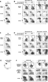
Ly6CHi-CD11cHi cells appear in the spleens of mice during CD40-mediated inflammatory responses. A. Gating scheme for identification of spleen subsets by flow cytometry showing the Ly6C v. CD11c plots used for the identification of Ly6CHi monocytes, Ly6CNeg monocytes, and CD11b+ DCs. B. Mice were injected with 100 μg of anti-CD40 mAb and spleen monocytes were analyzed at various time points post-injection. C. Identification of four populations in mice treated with anti-CD40 for 40hrs D. Plots shown in B were gated on Ly6CHi cells (solid line) for analysis of CD11c expression levels over time, and compared to the levels expressed by cDCs (dotted line) from the same mouse. Data are representative of 5 experiments, three mice per group. Results expressed as mean ± standard deviation from the mean.
Figure 2
Figure 2
Despite their high expression of CD11c, Ly6CHi-CD11cHi cells maintain a surface phenotype similar to activated Ly6CHi monocytes. A. Comparison of the surface expression of MHC and co-stimulatory molecules on Ly6CHi monocytes, Ly6CHi-CD11cHi cells, and CD11b+ cDCs from the spleen of the same mouse 40 hours after anti-CD40 treatment. Populations were identified as shown in figure 1D. B. Similar to A, surface phenotype of Ly6CNeg monocytes was analyzed in spleens of either control-treated mice or in mice treated with anti-CD40 for 40 or 90 hrs. Also shown is the surface phenotype of Ly6CHi-CD11cHi cells from the mice treated with anti-CD40 for 40h. In all cases, the indicated surface marker staining (solid line) was compared to staining obtained with an isotype control (dashed line). Data are representative of 4 experiments, two mice per group.
Figure 3
Figure 3. Ly6CHi-CD11cHi cells arise from monocytes in vivo
A.Mice were injected with 1.0-μm YG beads and 1 hour later, cells that had internalized beads (mostly monocytes) were analyzed and overlaid onto all cells. B. Mice were injected with 1.0-μm YG beads, and then either control-treated, or injected with anti-CD40 mAb. To track the fate of the cells that had internalized beads, 24 or 40 hours later, the phenotype of the bead containing cells were analyzed and overlaid onto all the cells. C. Similar to A, but cells were examined 24 hours after bead injection. D. Similar to B, but mice were injected with anti-CD40 24 hours after injection of beads. E. Spleen Ly6CHi monocytes were sorted to purity from CD45.1 mice using the gates shown in Figure 1. F. ~5×10⁵ purified monocytes as shown in E were injected into CD45.2 mice, that were either control treated, or immediately injected with anti-CD40 mAb. 24 or 40 hours later, the phenotype of the endogenous and transferred spleen monocytes was analyzed by flow cytometry. Data are representative of 3 experiments, two mice per group.
Figure 4
Figure 4. Ly6CHi-CD11cHi cells retain the scavenging capacity of monocytes
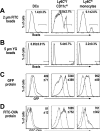
A. Comparison of the capacity of Ly6CHi monocytes, Ly6CHi-CD11cHi cells, and CD11b+ cDCs to internalize particulate antigens. Mice that had been treated with anti-CD40 mAb 24 hours earlier were injected with 2.0 μm fluorescent particles, and 60 minutes later, bead capture by splenocytes was analyzed by flow cytometry. Percentages shown represent the percent of cells that internalized one or more beads. B. Similar to A, but 5.0 μm particles were used. C. Similar to A and B but mice were injected with soluble GFP protein. After 30 minutes, GFP capture was analyzed by comparing the florescence signal of cells from a mouse injected with GFP (solid line) as compared to a mouse similarly treated with anti-CD40 but not injected with GFP (dashed line). Spleen populations were gated as described in Figure 1. The insert in each panel shows the MFI difference between GFP-injected mice and control mice is shown ± standard error. D. The same experimental approach as in C, but mice were injected intravenously with FITC-OVA (instead of GFP as used in C). Data are representative of three experiments, three mice per group. Results are expressed as mean ± standard deviation from the mean.
Figure 5
Figure 5. Ly6CHi-CD11cHi cells retain functional properties of monocytes
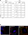
A. Despite a large capacity for antigen capture, Ly6CHi-CD11cHi cells do not present the internalized antigen to T-cells, a feature similar to monocytes but in contrast to dendritic cells. Mice were treated with anti-CD40 and 40 later injected intravenously with 1mg OVA protein. 30 minutes after OVA injection splenocytes were harvested and Ly6CHi-CD11cHi cells, Ly6CHi monocytes, and CD11bHi-CD11cHi cDCs were isolated from the same spleen and separately co-cultured with CFSE-labeled OT-I or OT-II T-cells over a range of T-cell/DC ratios. After 60h, antigen presentation was assessed by flow cytometric analysis of CFSE0-dilution to measure T-cell proliferation. B. Distribution of cDC and monocyte in the spleen of untreated control mice vs. anti-CD40 treated mice. A significant portion of the cDCs (Ly6CNeg-CD11cHi) are found in the T-cell zone after treatment with anti-CD40, while all Ly6CHi cells remain outside the white pulp. Data are representative of 3 independent experiments two mice per group.
Figure 6
Figure 6. Similar to Ly6CHi monocytes, Ly6CHi-CD11cHi cells ultimately differentiate into Ly6CNeg monocytes
A. The disappearance of Ly6CHi-CD11cHi cells 90 hours after anti-CD40 treatment, correlates with an increase in Ly6CNeg monocytes. B. Similar to Figure 3B, purified CD45.1 monocytes were injected into CD45.2 mice, which were immediately injected with anti-CD40 mAb. 90 hours later, the phenotype of the endogenous and transferred spleen monocytes was analyzed by flow cytometry. C. Similar to figure 3F, mice were injected with 1.0-μm YG beads, and then immediately injected with anti-CD40 mAb. 90 hours later the phenotype of the bead containing cells were analyzed and overlaid over all the cells. Data are representative of 3 experiments, two mice per group.

References

    1. French RR, Chan HT, Tutt AL, Glennie MJ. CD40 antibody evokes a cytotoxic T-cell response that eradicates lymphoma and bypasses T-cell help. Nat Med. 1999;5:548–553. - PubMed
    1. Lefrancois L, Altman JD, Williams K, Olson S. Soluble antigen and CD40 triggering are sufficient to induce primary and memory cytotoxic T cells. J Immunol. 2000;164:725–732. - PubMed
    1. Clarke SR. The critical role of CD40/CD40L in the CD4-dependent generation of CD8+ T cell immunity. Journal of leukocyte biology. 2000;67:607–614. - PubMed
    1. Hawiger D, Inaba K, Dorsett Y, Guo M, Mahnke K, Rivera M, Ravetch JV, Steinman RM, Nussenzweig MC. Dendritic cells induce peripheral T cell unresponsiveness under steady state conditions in vivo. J Exp Med. 2001;194:769–779. - PMC - PubMed
    1. Bonifaz L, Bonnyay D, Mahnke K, Rivera M, Nussenzweig MC, Steinman RM. Efficient targeting of protein antigen to the dendritic cell receptor DEC-205 in the steady state leads to antigen presentation on major histocompatibility complex class I products and peripheral CD8+ T cell tolerance. J Exp Med. 2002;196:1627–1638. - PMC - PubMed

Publication types

MeSH terms