Photoacoustic tomography: in vivo imaging from organelles to organs
- PMID: 22442475
- PMCID: PMC3322413
- DOI: 10.1126/science.1216210
Photoacoustic tomography: in vivo imaging from organelles to organs
Abstract
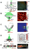
Photoacoustic tomography (PAT) can create multiscale multicontrast images of living biological structures ranging from organelles to organs. This emerging technology overcomes the high degree of scattering of optical photons in biological tissue by making use of the photoacoustic effect. Light absorption by molecules creates a thermally induced pressure jump that launches ultrasonic waves, which are received by acoustic detectors to form images. Different implementations of PAT allow the spatial resolution to be scaled with the desired imaging depth in tissue while a high depth-to-resolution ratio is maintained. As a rule of thumb, the achievable spatial resolution is on the order of 1/200 of the desired imaging depth, which can reach up to 7 centimeters. PAT provides anatomical, functional, metabolic, molecular, and genetic contrasts of vasculature, hemodynamics, oxygen metabolism, biomarkers, and gene expression. We review the state of the art of PAT for both biological and clinical studies and discuss future prospects.
Figures


References
-
- Wang LV, Wu H. Biomedical Optics: Principles and Imaging. Wiley; Hoboken, NJ: 2007.
-
-
The optical diffusion limit represents the depth of the quasi-ballistic regime in biological tissue beyond which light propagating along the predefined linear trajectory becomes too weak to be detected in practice. It is usually equated with the transport mean free path, i.e., the mean distance between two consecutive equivalent isotropic scattering events.
-
-
- Culver JP, Ntziachristos V, Holboke MJ, Yodh AG. Opt Lett. 2001;26:701. - PubMed
-
-
The sensitivity is defined here as the ratio of the fractional change in the photoacoustic signal to the fractional change in the optical absorption coefficient.
-
Publication types
MeSH terms
Grants and funding
LinkOut - more resources
Full Text Sources
Other Literature Sources
Research Materials

