. 2012 Jun;107(12):3235-45.
doi: 10.1152/jn.00501.2011. Epub 2012 Mar 21.

Theoretical principles underlying optical stimulation of a channelrhodopsin-2 positive pyramidal neuron

Affiliations

Theoretical principles underlying optical stimulation of a channelrhodopsin-2 positive pyramidal neuron

Thomas J Foutz et al. J Neurophysiol. 2012 Jun.

Abstract

Optogenetics is an emerging field of neuromodulation that permits scaled, millisecond temporal control of the membrane dynamics of genetically targeted cells using light. Optogenetic technology has revolutionized neuroscience research; however, numerous biophysical questions remain on the optical and neuronal factors impacting the modulation of neural activity with photon-sensitive ion channels. To begin to address such questions, we developed a computational tool to explore the underlying principles of optogenetic neural stimulation. This "light-neuron" model consists of theoretical representations of the light dynamics generated by a fiber optic in brain tissue, coupled to a multicompartment cable model of a cortical pyramidal neuron embedded with channelrhodopsin-2 (ChR2) membrane dynamics. Simulations revealed that the large energies required to generate an action potential are primarily due to the limited conductivity of ChR2, and that the major determinants of stimulation threshold are the surface area of illuminated cell membrane and proximity to the light source. Our results predict that the activation threshold is sensitive to many of the properties of ChR2 (density, conductivity, and kinetics), tissue medium (scattering and absorbance), and the fiber-optic light source (diameter and numerical aperture). We also illustrate the impact of redistributing the ChR2 expression density (uniform vs. nonuniform) on the activation threshold. The model system developed in this study represents a scientific instrument to characterize the effects of optogenetic neuromodulation, as well as an engineering design tool to help guide future development of optogenetic technology.


Figures

Fig. 1.
Fig. 1.
Light-neuron model. A: blue light is absorbed by channelrhodopsin-2 (ChR2), followed by a conformational change, opening the channel for the passage of cations. ChR2 structure image was adapted from Müller et al. (2011). B: representation of a fiber-optic illuminating a layer V pyramidal neuron. C: neuron represented by a multicompartment cable model. ChR2 is only activated in compartments directly illuminated by the light source. D: ChR2 is modeled in four states, with two open and two closed states. Channels can transition between states with rate constants as described by Nikolic et al. (2009). e12, e21, Kd1, Kd2, Kr: rate constants. E: electrical circuit representation of the soma-dendritic compartments. Na+, Na+1.2, Na+1.6: sodium channels; K+, potassium channel; Kslow+, slow, noninactivating potassium channel; KCa+, calcium-dependent potassium channel; Ca2+, high-voltage-activated calcium channel, with decay of internal calcium concentration; Leak, passive leakage channel; Cm, membrane capacitance.
Fig. 2.
Fig. 2.
Fiber-optic light model. A: three-dimensional representation of fiber-optic light output. Lossless demonstrates Gaussian-distributed light in a vacuum, with no geometric spread. Geometric spread and scattering are shown independently and then combined. B and C: light transmission and irradiance as a function of the fiber-optic-to-soma distance. The fiber optic was directed at the soma from distances ranging 0.1 to 2.0 mm (50 μm resolution). Optical stimuli were applied with 5-ms duration. Three different fiber-optic diameters (0.1, 0.2, and 0.4 mm) were simulated. B: transmission of light through diffuse, scattering tissue. Transmission is a measure of the drop in irradiance, due to conical geometry, absorbance, and scattering of emitted light. Comparison to results are reported in rodent brain tissue with 0.4-mm-diameter fiber optic (▴) (Gradinaru et al. 2009). I, light at each point in space; I0, center of fiber-optic output. C: threshold source light irradiance required to generate a propagating action potential in a layer V pyramidal neuron with a fiber-optic-neuron orientation depicted in Fig. 1B.
Fig. 3.
Fig. 3.
Strength-duration relationships. The fiber-optic-neuron orientation was as shown in Fig. 1B. Irradiance required to generate an action potential was determined for 10-μs to 10-ms pulse widths. Fiber-optic diameter, 0.2 mm. Fiber-optic-to-soma distances were 0.5, 1, and 1.5 mm.
Fig. 4.
Fig. 4.
Repetitive stimulation. Neuron firing frequency rates are shown in response to optical stimulation with pulse frequencies ranging from 1 to 200 Hz. Top: response at different stimulation irradiance levels: 100%, 120%, and 140% of single pulse action potential threshold (Ithres). Bottom: response with different ChR2 channel densities: 75%, 100%, and 150% of our default density (ρChR2). Irradiance level was 120% of single spike threshold.
Fig. 5.
Fig. 5.
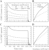
Response of ChR2 channels. Membrane voltage was clamped to −70 mV. Fiber-optic-neuron orientation is as shown in Fig. 1B. A: total ChR2 current induced in the neuron with irradiance ranging from 10 to 500 mW/mm2 for 1 s. B: peak and plateau ChR2 currents, both of which increase for larger irradiance. C: percentage of all ChR2 channels opened by given irradiance levels. D: peak and plateau percentage of channels opened, both of which increase with larger irradiance levels.
Fig. 6.
Fig. 6.
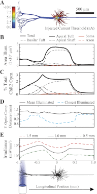
Optical stimulation profile. A: current injection threshold at each individual model compartment required to generate an action potential in a nonilluminated neuron. 500-μm scale bar is valid for all x-axes. B–D: fiber optic is oriented perpendicular to the long axis of the neuron (1-mm fiber-optic-to-neuron distance), illuminating the neuron at 40-μm intervals along the longitudinal position of the neuron. Stimulations were performed with a 0.2-mm-diameter fiber optic, and 5-ms stimulus duration. The threshold irradiance was calculated for each longitudinal position. B: area of membrane illuminated (transmittance > 0.001). C: percentage of total ChR2 channels in the open state at the end of a threshold stimulation pulse. D: distance from the output end of the fiber optic to either the closest illuminated neuron compartment, or the average position of all illuminated compartments (transmittance > 0.001). E: threshold irradiance level (±0.1%) required to generate a propagating action potential from different fiber-optic-to-neuron distances.
Fig. 7.
Fig. 7.
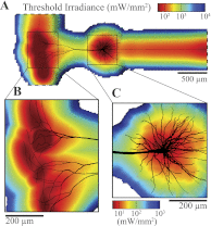
Threshold irradiance contours. Color represents light irradiance threshold (mW/mm2) required to generate a propagating action potential (±0.1%). Simulations were performed with 0.2-mm-diameter fiber optic and stimulus duration of 5 ms. A: threshold for action potential generation at over 8,000 different fiber-optic-to-neuron positions with the fiber optic incrementally moved in 10-μm steps in a plane 500 μm above the long axis of the neuron. B and C: detail views of stimulation from a plane 100 μm above the neuron. Threshold irradiance color bar in C also applies to B.
Fig. 8.
Fig. 8.
Cortical stimulation. A and B: artificial rendering of light-neuron model integration with a Thy1-ChR2 transgenic mouse line 18 (Wang et al. 2007) (coronal histological slice image provided by Nicholas Chow and George Augustine). B: close-up of cortical layers, with model neurons overlaid in the layer V region and fiber optic oriented perpendicular to the surface of the cortex. C: irradiance threshold to generate a propagating action in a population of neurons. The fiber-optic tip was 0.5 mm from the apical tuft of the closest neuron (1.5 mm from the soma of the closest neuron). Neuron color corresponds to the irradiance threshold.
Fig. 9.
Fig. 9.
Parameter sensitivity analysis. Each model parameter was varied independently (±50%), and the threshold irradiance level necessary to generate an action potential was determined (±0.1%). Default values are listed in Table 1.
Fig. 10.
Fig. 10.
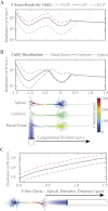
Variable ChR2 expression. A and B: the fiber optic was placed perpendicular to long axis of the neuron (1-mm distance), and the threshold irradiance required to generate an action potential was calculated (±0.1%). A: stimulation profile of a neuron with 5, 10, or 50 million channels distributed uniformly in the soma-dendritic compartments of the model. ChR2 expression in the axon was uniform and constant at the default model value. B: stimulation profile with 3 different distributions of 10 million ChR2 channels: basal/soma with higher expression of ChR2 in the basal tuft and somatic compartments, uniform with equal channel density across all compartments, and apical with higher expression of ChR2 in the apical tuft. C: the fiber optic was placed along the longitudinal axis of the neuron, directed at the apical tuft. The threshold irradiance was calculated as a function of the fiber-optic distance from the apical tuft for the 3 different ChR2 distributions used in B.

References

    1. Aravanis AM, Wang LP, Zhang F, Meltzer LA, Mogri MZ, Schneider MB, Deisseroth K. An optical neural interface: in vivo control of rodent motor cortex with integrated fiberoptic and optogenetic technology. J Neural Eng 4: S143–S156, 2007 - PubMed
    1. Berndt A, Prigge M, Gradmann D, Hegemann P. Two open states with progressive proton selectivities in the branched channelrhodopsin-2 photocycle. Biophys J 98: 753–761, 2010 - PMC - PubMed
    1. Berndt A, Schoenenberger P, Mattis J, Tye KM, Deisseroth K, Hegemann P, Oertner TG. High-efficiency channelrhodopsins for fast neuronal stimulation at low light levels. Proc Natl Acad Sci USA 108: 7595–7600, 2011 - PMC - PubMed
    1. Carnevale NT, Hines ML. The NEURON Book (1st Ed.). Cambridge, UK: Cambridge University Press, 2009
    1. Deisseroth K, Feng G, Majewska A, Miesenbock G, Ting A, Schnitzer M. Next-generation optical technologies for illuminating genetically targeted brain circuits. J Neurosci 26: 10380–10386, 2006 - PMC - PubMed

Publication types

LinkOut - more resources