. 2012 Jun;107(12):3342-8.
doi: 10.1152/jn.00746.2011. Epub 2012 Mar 21.

The relative importance of retinal error and prediction in saccadic adaptation

Affiliations

The relative importance of retinal error and prediction in saccadic adaptation

Thérèse Collins et al. J Neurophysiol. 2012 Jun.

Abstract

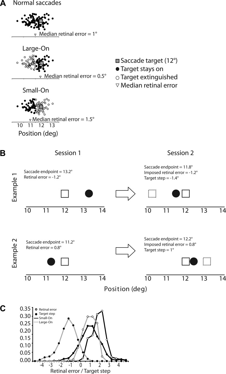
When saccades systematically miss their visual target, their amplitude adjusts, causing the position errors to be progressively reduced. Conventionally, this adaptation is viewed as driven by retinal error (the distance between primary saccade endpoint and visual target). Recent work suggests that the oculomotor system is informed about where the eye lands; thus not all "retinal error" is unexpected. The present study compared two error signals that may drive saccade adaptation: retinal error and prediction error (the difference between predicted and actual postsaccadic images). Subjects made saccades to a visual target in two successive sessions. In the first session, the target was extinguished during saccade execution if the amplitude was smaller (or, in other experiments, greater) than the running median, thereby modifying the average retinal error subjects experienced without moving the target during the saccade as in conventional adaptation paradigms. In the second session, targets were extinguished at the start of saccades and turned back on at a position that reproduced the trial-by-trial retinal error recorded in the first session. Despite the retinal error in the first and second sessions having been identical, adaptation was severalfold greater in the second session, when the predicted target position had been changed. These results argue that the eye knows where it lands and where it expects the target to be, and that deviations from this prediction drive saccade adaptation more strongly than retinal error alone.


Figures

Fig. 1.
Fig. 1.
A: schematic illustration of amplitude-dependent visual feedback. In normal saccades (top), endpoints tend to undershoot the visual target. Thus the median retinal error tends to be positive. In the “Large-On” condition (middle), the visual target was available after the primary saccade only when its amplitude was larger than the median. Thus the distribution of retinal errors experienced by the subject was smaller than usual. In the “Small-On” condition (bottom), the visual target was available after the primary saccade only when its amplitude was smaller than the median. The distribution of retinal errors experienced by the subject was larger than usual. B: 2 examples of corresponding trials during the 2 sessions. The saccade target (open square) was always at 12°. In the first example (top), in session 1 the saccade endpoint (black dot) on trial n was 13.2°, which corresponds to a retinal error of −1.2°. In session 2, the saccade endpoint on trial n was 11.8°. To obtain a retinal error of −1.2°, the target had to be stepped back to position 10.6° (dashed square), a step of −1.4°. In the second example (bottom), in session 1 the saccade endpoint was 11.2° and retinal error was 0.8°. In session 2, since the endpoint was 12.2°, obtaining a retinal error of 0.8° required stepping the target forward by 1°. C: distribution of retinal errors and target steps in Large-On and Small-On conditions, pooled over all subjects. The zero mark on the x-axis corresponds to the target location; thus a step of 0 means that the target did not step, and a retinal error of 0 means that the saccade endpoint was at the same location as the postsaccadic target.
Fig. 2.
Fig. 2.
Example time courses of adaptation (amplitude as a function of trial). Each point is 1 saccade. Larger symbols with standard deviation error bars on left and right correspond to the mean endpoint in baseline and posttest epochs, respectively. Curves represent the sliding median (n = 50). Recall that in session 1 only saccades below (in the Small-On condition) or above (in the Large-On condition) this median received visual feedback. Note that at trial ∼400 in the Large-On condition (upper circled data point) saccade amplitude in session 1 was 13.1°; because the target was at 12°, this resulted in a retinal error of −1.1°. In session 2 at the same trial number (lower circled data point), the saccade amplitude was 8.7°, so we moved the target to 7.6° to replicate the retinal error seen on session 1.
Fig. 3.
Fig. 3.
Percent amplitude change over the course of adaptation in Large-On (left) and Small-On (right) conditions. The data plotted are the difference between the posttest and baseline open-loop trials (target extinguished upon saccade onset and no reappearance). Gray and white bars are individual subjects; black bars are means ± SE. Percent amplitude change was calculated as % amplitude change = (posttest amplitude − baseline amplitude)/baseline amplitude.

References

    1. Abel L, Schmidt D, Dell'Osso L, Daroff R. Saccadic system plasticity in humans. Ann Neurol 4: 313–318, 1978 - PubMed
    1. Bahcall D, Kowler E. Illusory shifts in visual direction accompany adaptation of saccadic eye movements. Nature 26: 864–866, 1999 - PubMed
    1. Bahcall D, Kowler E. The control of saccadic adaptation: implications for the scanning of natural visual scenes. Vision Res 40: 2779–2796, 2000 - PubMed
    1. Blakemore SJ, Wolpert DM, Frith CD. Why can't you tickle yourself? Neuroreport 11: 11–15, 2000 - PubMed
    1. Bock O, Goltz H, Bélange S, Steinback M. On the role of extraretinal signals for saccade generation. Exp Brain Res 104: 349, 1995 - PubMed

Publication types

MeSH terms

LinkOut - more resources