. 2012;7(3):e33420.
doi: 10.1371/journal.pone.0033420. Epub 2012 Mar 19.

Mechanism of RPE cell death in α-crystallin deficient mice: a novel and critical role for MRP1-mediated GSH efflux

Affiliations

Mechanism of RPE cell death in α-crystallin deficient mice: a novel and critical role for MRP1-mediated GSH efflux

Parameswaran G Sreekumar et al. PLoS One. 2012.

Abstract

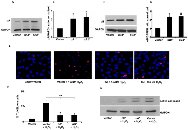
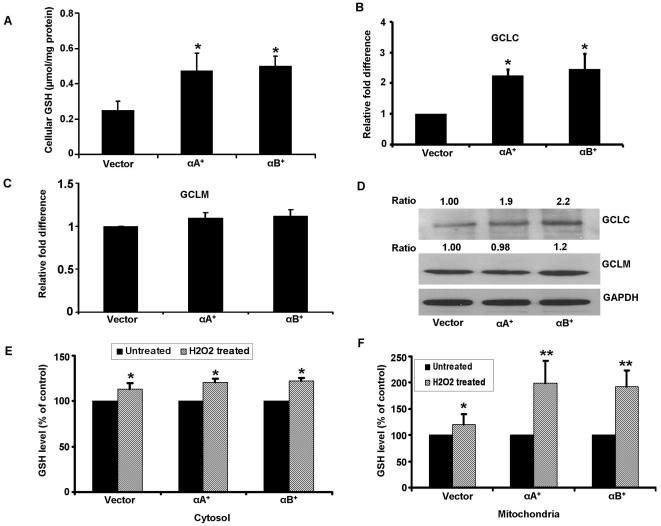
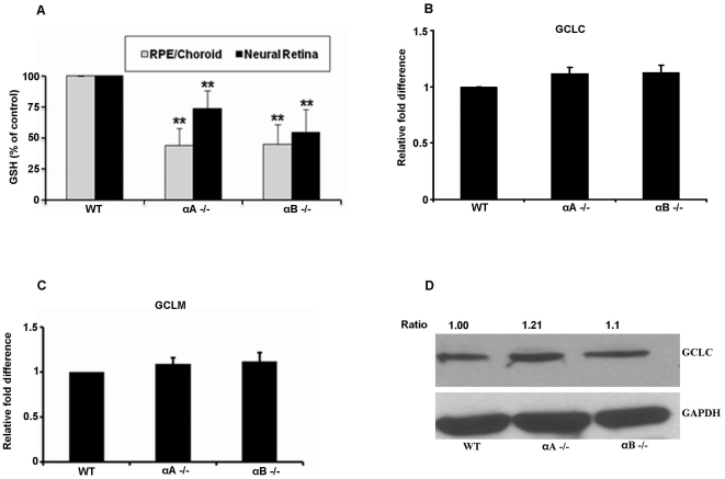
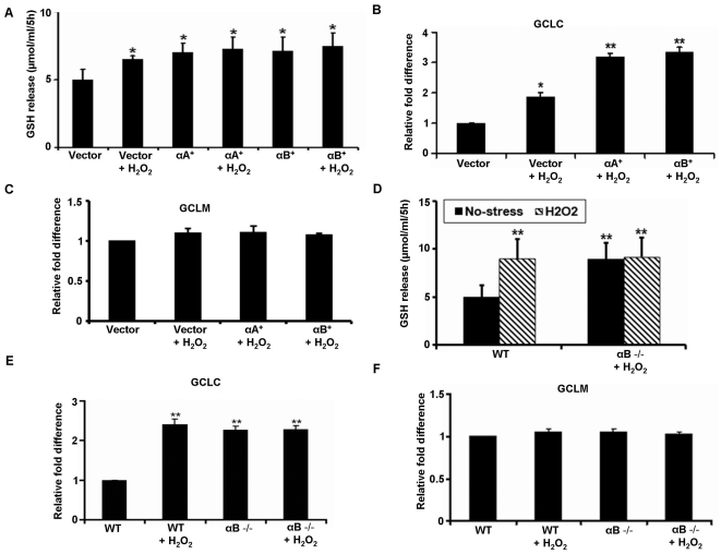
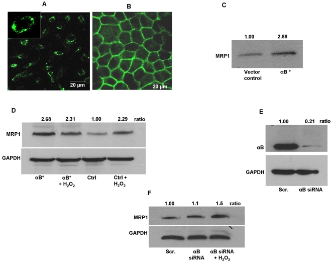
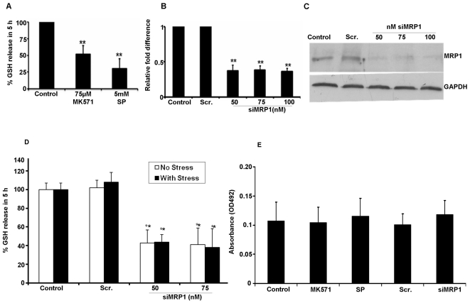
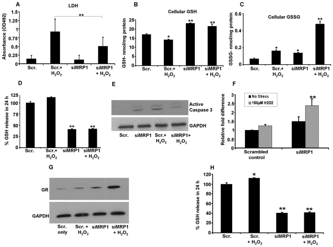
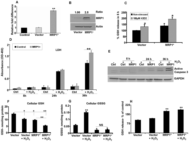
Absence of α-crystallins (αA and αB) in retinal pigment epithelial (RPE) cells renders them susceptible to oxidant-induced cell death. We tested the hypothesis that the protective effect of α-crystallin is mediated by changes in cellular glutathione (GSH) and elucidated the mechanism of GSH efflux. In α-crystallin overexpressing cells resistant to cell death, cellular GSH was >2 fold higher than vector control cells and this increase was seen particularly in mitochondria. The high GSH levels associated with α-crystallin overexpression were due to increased GSH biosynthesis. On the other hand, cellular GSH was decreased by 50% in murine retina lacking αA or αB crystallin. Multiple multidrug resistance protein (MRP) family isoforms were expressed in RPE, among which MRP1 was the most abundant. MRP1 was localized to the plasma membrane and inhibition of MRP1 markedly decreased GSH efflux. MRP1-suppressed cells were resistant to cell death and contained elevated intracellular GSH and GSSG. Increased GSH in MRP1-supressed cells resulted from a higher conversion of GSSG to GSH by glutathione reductase. In contrast, GSH efflux was significantly higher in MRP1 overexpressing RPE cells which also contained lower levels of cellular GSH and GSSG. Oxidative stress further increased GSH efflux with a decrease in cellular GSH and rendered cells apoptosis-prone. In conclusion, our data reveal for the first time that 1) MRP1 mediates GSH and GSSG efflux in RPE cells; 2) MRP1 inhibition renders RPE cells resistant to oxidative stress-induced cell death while MRP1 overexpression makes them susceptible and 3) the antiapoptotic function of α-crystallin in oxidatively stressed cells is mediated in part by GSH and MRP1. Our findings suggest that MRP1 and α crystallin are potential therapeutic targets in pathological retinal degenerative disorders linked to oxidative stress.


Conflict of interest statement

Competing Interests: The authors have declared that no competing interests exist.

Figures

Figure 1
Figure 1. αA and αB crystallin protect RPE cells from H2O2-induced oxidative stress.
αA and αB crystallin expression in two stably overexpressing ARPE-19 cell lines (αA1+, αA2+, αB1+, αB2+) was significantly higher than empty vector controls (A–D). Immunoblot analysis of ARPE-19 cells expressing αA crystallin and αB crystallin (A,C). Protein samples (25–75 µg) were separated by 15% Tris-HCL gel, transferred to PVDF membrane, and subsequently reacted with rabbit αA crystallin and αB crystallin antibodies. Protein expression quantified by densitometry is shown as ratio normalized with GAPDH (B, D). Cell death induced by H2O2 was significantly reduced in α-crystallin overexpressing ARPE-19 cells (E). α-Crystallin overexpressing cells were incubated with 150 µM H2O2 for 24 h in serum free medium and cell death was analyzed by TUNEL staining and semi-quantification of TUNEL positive cells are presented as percentage of dead cells in F. Blue: DAPI nuclear staining; Red: TUNEL positive cells. Caspase 3 activation with H2O2 in α-crystallin overexpressing cells (G). Twenty four hours after treatment, cell lysates (60 µg total protein) were run in inmmunoblots for active caspase 3 and then reprobed for GAPDH as a loading control. αA+ -αA crystallin overexpressing clones, αB+ -αB crystallin overexpressing clones, αA- αA crystallin, αB- αB crystallin. ** P<0.01 H2O2 vs αA+ and αB+. * P<0.05 Vector vs α-crystallin clones.
Figure 2
Figure 2. Intracellular GSH levels in α-crystallin overexpressing cells.
Cellular GSH levels in ARPE-19 cells stably overexpressing αA crystallin and αB crystallin increased significantly (P<0.05) when compared to vector only cells (A). Relative expression of the mRNA levels of the catalytic unit (GCLC) and modifier unit (GCLM) of GSH biosynthetic enzyme GCL (B, C). A significant increase in GCLC was found for mRNA and protein while no significant change was observed either at the gene or protein levels for the modifier unit (B–D). Protein bands were quantified and presented as ratio normalized to loading control, GAPDH. Panels E and F show GSH levels in the cytosol and mitochondrial fractions from vector control and α-crystallin overexpressing ARPE-19 cells challenged with 150 µM H2O2 for 24 h. While the magnitude of increase in the cytosolic pool is relatively smaller, a significant (P<0.01) increase in mitochondrial pool of GSH was observed in α-crystallin overexpressing cells. Data are normalized to control taken as 100%. *P<0.05 vs controls; ** P<0.01 vs controls.
Figure 3
Figure 3. Cellular GSH levels in the αA and αB crystallin knockout (KO) mouse retina.
(A) GSH levels in two tissue segments of the eye, namely RPE/Choroid and neural retina. Data, presented as percentage over control age-matched mice, showed a significant 50% decrease in GSH level in the neural retina and a ∼25–30% decrease in the RPE/choroid in αA crystallin KO (αA −/−) and αB crystallin KO (αB −/−) samples. RNA and protein were extracted from the posterior eye cup of the crystallin KO and WT mice. The gene expression of the catalytic (B) and the modifier unit (C) as well as the protein expression (D) of the GSH rate limiting enzyme GCLC did not show any significant change among αA (−/−), αB(−/−) and WT mice. GCLC- glutamate-cysteine ligase, catalytic subunit, GCLM- glutamate-cysteine ligase, modifier subunit, KO- knock out, WT- wild type. ** P<0.01 vs controls.
Figure 4
Figure 4. GSH export from H2O2-treated α-crystallin overexpressing and αB crystallin KO RPE cells.
αA crystallin and αB crystallin overexpressing cells were treated with 150 µM H2O2 for 5 h in serum-free medium and extracellular accumulation of GSH was measured. Vector only cells treated in the same fashion served as controls (A). A significant (P<0.01 vs control) gene upregulation of GCLC (B) was observed while no significant change occurred in GCLM (C). GSH efflux in non-stressed RPE cells isolated from αB crystallin KO mice showed a significant increase when compared to RPE cells isolated from WT mice (D). However, oxidative stress (150 µM H2O2 for 5 h) resulted in a significant (P<0.01) increase in GSH efflux only in the WT RPE and no further significant increase in efflux was observed in RPE from αB crystallin KO mice. H2O2-induced stress produced a significant upregulation of the GCLC (E) while GCLM did not show any significant change (F). GCLC- glutamate-cysteine ligase, catalytic subunit, GCLM- glutamate-cysteine ligase, modifier subunit, WT- wild type. *P<0.05, ** P<0.01.
Figure 5
Figure 5. MRP1 is localized to the plasma membrane in human RPE cells.
ARPE-19 cells (A) and polarized RPE monolayer from fetal human RPE cells (B) were fixed and incubated with a monoclonal antibody against MRP1 followed by a fluorescence labeled anti-mouse secondary antibody. Images were taken on a confocal laser-scanning microscope. (C) Vector control and αB crystallin overexpressing cells were labeled with biotin and surface labeled protein samples were analyzed by immunoblot for MRP1. A significant almost 3 fold increase in MRP1 protein expression was found. (D) MRP1 protein expression in vector control and αB crystallin overexpressing cells with and without oxidative stress. Cells were incubated with 300 µM H2O2 for 36 h in serum-free medium and total cell lysate was subjected to immunoblot analysis for MRP1 protein. Expression of MRP1 protein was >2 fold in αB crystallin overexpressing cells as compared to vector control cells. Treatment with H2O2 did not significantly alter MRP1 expression in αB crystallin overexpressing cells, however, a significant >2-fold increase was observed in vector control cells exposed to H2O2. (E) ARPE-19 cells were transiently transfected with scrambled and αB crystallin siRNA (50 nM) and protein was harvested 72 h post transfection. Whole cell lysates (20 µg of total protein) were subjected to immunoblot analysis using a rabbit polyclonal antibody against αB crystallin. αB crystallin protein expression was markedly reduced by >80% in siRNA transfected cells 72 h post-transfection. (F) MRP1 expression in αB crystallin-silenced cells with and without oxidative stress with H2O2. Transfected cells were treated with 200 µM H2O2 for 24 h in serum free medium and total protein (100 µg) was subjected to immunoblot analysis. While there is no apparent change in MRP1 protein expression in αB crystallin-silenced cells, H2O2 increased MRP1 expression 1.5 fold in αB crystallin-silenced cells. GAPDH was used as a loading control for all immunoblot analyses. GAPDH- Glyceraldehyde 3-phosphate dehydrogenase, MRP1- Multidrug resistance protein 1, αB+- αB crystallin. Scr. -Scrambled.
Figure 6
Figure 6. Inhibition of MRP1 significantly decreases basal and apoptotic GSH efflux.
GSH efflux in ARPE-19 cells incubated with two MRP1 inhibitors (75 µM MK571 and 5 mM sulfinpyrazone [SP]) for 5 h (A). ARPE-19 cells were transfected with MRP1 siRNA at the indicated concentrations. RNA and protein were extracted 48 h post transfection. MRP1 mRNA and protein expression was significantly (P<0.01 vs scrambled control) decreased when compared to control or scrambled siRNA alone (B,C). GSH release from MRP1-silenced ARPE-19 cells incubated with 150 µM H2O2 for 5 h in serum free medium. A significant decrease (P<0.001 vs scrambled control) in GSH release was observed in 5 h. A decrease in GSH release only in control cells with no additional change in MRP1 inhibited cells was found and H2O2 treatment did not cause any further change (D). LDH release measured in MRP1 inhibited cells was not affected by any treatment (E). Values are mean± SE (n = 3–4). LDH- Lactate dehydrogenase. ** Significantly different from control cells, P<0.01.
Figure 7
Figure 7. MRP1-inhibited RPE cells are resistant to cell death.
MRP1 silenced and control cells were incubated with 150 µM H2O2 for 24 h in serum-free medium. 24 h LDH release was quantified in a 96 well plate reader. LDH release was significantly reduced in MRP1 silenced cells challenged with H2O2 when compared to similarly treated scrambled transfected cells (A). Cellular GSH (B) and GSSG (C) levels were significantly higher in MRP1 silenced cells when compared to scrambled controls. GSH efflux was inhibited by 60% in MRP1 silenced cells and did not change further with oxidant injury (D). Caspase activation, determined by immunoblot of active caspase 3 was reduced in MRP1-silenced cells challenged with H2O2 when compared to scrambled control cells (E). Glutathione reductase (GR) was significantly upregulated at the mRNA (F) and protein levels (G) in MRP1 silenced cells exposed to H2O2. Densitometric values are presented as ratio normalized to control (H). * P<0.05, ** P<0.01.
Figure 8
Figure 8. GSH efflux in MRP1 overexpressing ARPE-19 cells.
(A) Real-time PCR was performed from RNA extracted from control (parental cells), vector transfected cells and cells transfected with human MRP1. MRP1 mRNA levels were normalized to GAPDH and data are presented as relative fold change over control cells. (B) Immunoblot analysis for MRP1 protein expression. A significant 2.5 fold increase in MRP1 was found. (C) MRP1 overexpressing cells release significantly higher quantities of GSH when compared to control cells. Oxidative stress (150 µM H2O2 for 5 h) further increased GSH release from control as well as from MRP1 overexpressing cells. (D) LDH release was not significantly altered in cells challenged with H2O2 for 5 h in both control and in MRP1 overexpressing cells. However, LDH release showed a progressive increase when H2O2 exposure was extended to 24 h and 36 h (n = 9). (E) A time dependent activation of caspase 3 expression was found with H2O2 treatment and this increase was maximal at 36 h. Cellular GSH (F) and GSSG (G) levels were significantly decreased in MRP1 overexpressing cells and oxidative stress with H2O2 further decreased GSH levels. GSSG levels showed a significant increase in H2O2 treated vector control cells and were very low in MRP1 overexpressed cells in the presence or absence of H2O2. (H) GSH efflux was higher in MRP1 overexpressed cells and oxidative stress further increased this efflux. * P<0.05, ** P<0.01.

References

    1. Gehrs KM, Anderson DH, Johnson LV, Hageman GS. Age-related macular degeneration–emerging pathogenetic and therapeutic concepts. Ann Med. 2006;38:450–471. - PMC - PubMed
    1. Cai X, McGinnis JF. Oxidative stress: the achilles' heel of neurodegenerative diseases of the retina. Front Biosci. 2012;17:1976–1995. - PubMed
    1. Markovets AM, Saprunova VB, Zhdankina AA, Fursova AZh, Bakeeva LE, et al. Alterations of retinal pigment epithelium cause AMD like retinopathy in senescence-accelerated OXYS rats. Aging (Albany NY) 2011;3:44–54. - PMC - PubMed
    1. Zhu D, Wu J, Spee C, Ryan SJ, Hinton DR. BMP4 mediates oxidative stress-induced retinal pigment epithelial cell senescence and is overexpressed in age-related macular degeneration. J Biol Chem. 2009;284:9529–9539. - PMC - PubMed
    1. Zhu D, Deng X, Xu J, Hinton DR. What determines the switch between atrophic and neovascular forms of age related macular degeneration? - the role of BMP4-induced senescence. Aging (Albany NY) 2009;1:740–745. - PMC - PubMed

Publication types

MeSH terms

Substances

LinkOut - more resources