. 2012 Jul;18(13-14):1479-89.
doi: 10.1089/ten.TEA.2011.0325. Epub 2012 Jun 12.

Conditioned media from mesenchymal stem cells enhanced bone regeneration in rat calvarial bone defects

Affiliations

Conditioned media from mesenchymal stem cells enhanced bone regeneration in rat calvarial bone defects

Masashi Osugi et al. Tissue Eng Part A. 2012 Jul.

Abstract

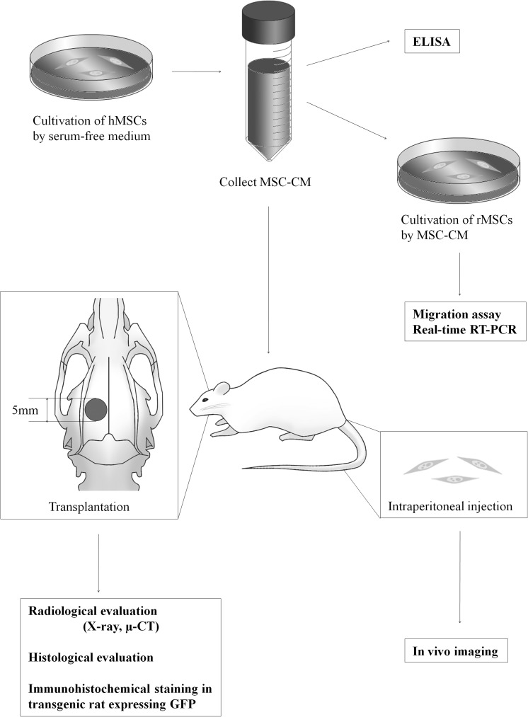
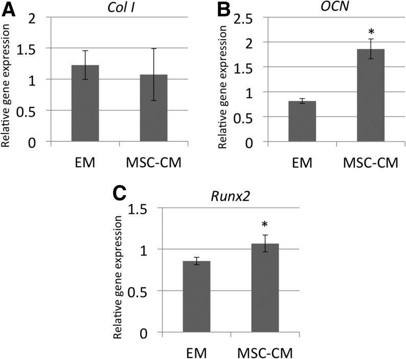
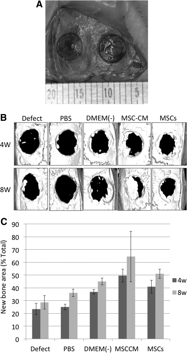
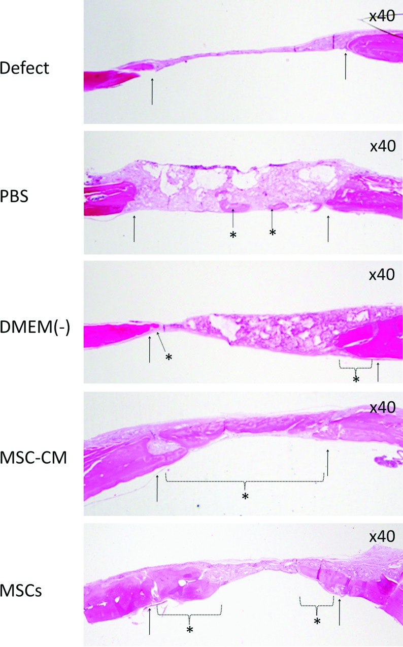
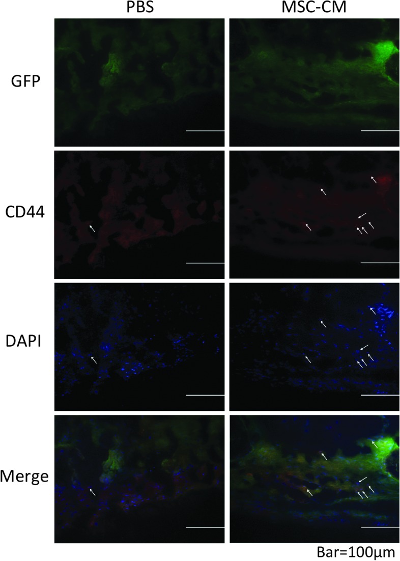
Tissue engineering has recently become available as a treatment procedure for bone augmentation. However, this procedure has several problems, such as high capital investment and expensive cell culture, complicated safety and quality management issues regarding cell handling, and patient problems with the invasive procedure of cell collection. Moreover, it was reported that stem cells secrete many growth factors and chemokines during their cultivation, which could affect cellular characteristics and behavior. This study investigated the effect of stem-cell-cultured conditioned media on bone regeneration. Cultured conditioned media from human bone marrow-derived mesenchymal stem cells (MSC-CM) enhanced the migration, proliferation, and expression of osteogenic marker genes, such as osteocalcin and Runx2, of rat MSCs (rMSCs) in vitro. MSC-CM includes cytokines such as insulin-like growth factor-1 and vascular endothelial growth factor. In vivo, a prepared bone defect of a rat calvarial model was implanted in five different rat groups using one of the following graft materials: human MSCs/agarose (MSCs), MSC-CM/agarose (MSC-CM), Dulbecco's modified Eagle's medium without serum [DMEM(-)]/agarose [DMEM(-)], PBS/agarose (PBS), and defect only (Defect). After 4 and 8 weeks, implant sections were evaluated using microcomputed tomography (micro-CT) and histological analysis. Micro-CT analysis indicated that the MSC-CM group had a greater area of newly regenerated bone compared with the other groups (p<0.05) and histological analysis at 8 weeks indicated that the newly regenerated bone bridge almost covered the defect. Interestingly, the effects of MSC-CM were stronger than those of the MSC group. In vivo imaging and immunohistochemical staining of transgenic rats expressing green fluorescent protein also showed that migration of rMSCs to the bone defect in the MSC-CM group was greater than in the other groups. These results demonstrated that MSC-CM can regenerate bone through mobilization of endogenous stem cells. The use of stem-cell-cultured conditioned media for bone regeneration is a unique concept that utilizes paracrine factors of stem cells without cell transplantation.


Figures

FIG. 1.
FIG. 1.
Outline of the experimental protocol. MSC-CM was collected after cultivation of hMSCs for 48 h in serum-free medium. A rat calvarial bone defect of 5 mm in diameter was produced in each parietal bone. The effect of MSC-CM on cell migration, proliferation, and gene expression in vitro, and of implanted MSC-CM on bone regeneration in vivo were then analyzed as shown. hMSCs, human mesenchymal stem cells; CM, conditioned media; rMSCs, rat MSCs; RT-PCR, reverse transcriptase-polymerase chain reaction.
FIG. 2.
FIG. 2.
Effect of MSC-CM on the migration and proliferation of rMSCs. (A) Transwell migration assay. The migration of rMSCs cultured in MSC-CM was enhanced compared with that of rMSCs cultured in DMEM(−). (B) BrdU cell proliferation assay. Proliferation of rMSCs was determined as the percentage of cells that incorporated BrdU. The proliferation of rMSCs was also enhanced when cultured with MSC-CM compared with culture in DMEM(−). Cells cultured in DMEM-30%FBS were used as a positive control for both (A) and (B). Asterisks indicate a significant difference between the indicated groups (*p<0.05). FBS, fetal bovine serum; BrdU, bromodeoxyuridine; rMSCs, rat MSCs; DMEM, Dulbecco's modified Eagle's medium.
FIG. 3.
FIG. 3.
Effect of MSC-CM on rMSC gene expression. The mRNA level of (A) Col I, (B) OCN, and (C) Runx2 genes in rMSCs cultured in MSC-CM or DMEM-10%FBS (EM) was assayed using real-time reverse transcriptase–polymerase chain reaction. Cells were lysed for extraction of total RNA on day 7 of culture in MSC-CM or EM, and equal amounts of total RNA (50 ng) were analyzed. The mRNA expression levels of Col I, OCN, and Runx2 were determined relative to the level of GAPDH mRNA in each sample and were quantified by calculation based on their standard curves as described in the Materials and Methods section. To quantitatively compare the levels of gene expression of the different samples, the expression coefficient for each mRNA on the ordinate was calculated by dividing the absolute level of expression of each mRNA (Col I, OCN, and Runx2) with the absolute level of expression of GAPDH mRNA in each sample. Each point represents the mean value calculated from five independent replicates, in which the difference was <10%. An asterisk indicates a significant difference between the EM and MSC-CM groups for the indicated gene (*p<0.05). Col I, collagen type I alpha 2; OCN, osteocalcin; EM, expansion medium.
FIG. 4.
FIG. 4.
Micro-CT analysis of bone regeneration following implantation of MSC-CM or controls into a bone defect. (A) Rat calvarial bone defects before implantation of the materials. A bone defect of 5 mm in diameter was prepared in each parietal bone. (B) Micro-CT images of the calvaria 4 and 8 weeks after implantation of the indicated materials. In the PBS, DMEM(−), MSC-CM, and MSCs groups, the materials were implanted as a mixture with an agarose gel, while in the Defect group, the defect was left unfilled. (C) The area of newly regenerated bone in each defect 4 and 8 weeks after implantation of the materials. The area of the newly regenerated bone (mm2) was calculated for each of the experimental groups. The regenerated area is expressed as a percentage of the entire defect area. There were statistical differences between the area of the MSC-CM and that of the other groups both at 4 and 8 weeks except between the MSC-CM and MSCs groups at 8 weeks (p<0.05). Micro-CT, microcomputed tomography; PBS, phosphate-buffered saline.
FIG. 5.
FIG. 5.
Histological analysis of newly regenerated bone after implantation. Newly regenerated bone in the defect (Defect), PBS/agarose (PBS), DMEM(−)/agarose [DMEM(−)], MSC-CM/agarose (MSC-CM), and hMSCs/agarose (MSCs) groups was evaluated histologically. Hematoxylin and eosin staining of calvarial histological sections was performed 8 weeks after implantation. The arrows indicate the edges of the host bone and the dotted arrow and dotted line with asterisks indicate the newly regenerated bone. The bone bridge almost covered the defect in the MSC-CM group. In the MSCs group, partial regenerated bone was observed. Agarose gel remnants were not seen in these two groups. In the PBS and DMEM(−) groups, some newly regenerated bone and remnants of the agarose gel were seen, while in the Defect group most of the defect was filled with connective tissue. Inflammatory responses were not observed in any group. Color images available online at www.liebertonline.com/tea
FIG. 6.
FIG. 6.
In vivo imaging of injected rMSC migration to implants. In vivo imaging analysis shows that DiR-labeled rMSCs that were injected into the caudal vein just after implantation of the materials into the calvarial bone defects started to migrate immediately after injection. At 1 h, 24 h, 48 h, and 1 week after injection, the signal of the fluorescent-labeled rMSCs was only detected in the tail and abdominal region in the control Defect group. We confirmed that there was no signal at the defect area in the cranium at each time point. In the PBS group, fluorescent signals were observed in the tail and the abdominal area at 24 and 48 h after injection, and very low signals were observed in the breast and the cranial area after 1 week. In the MSC-CM group, a moderate increase in signal intensity was observed in the area of the tail and in the abdominal region during the first 24 h after injection. Forty-eight hours after injection, the MSC-CM-implanted area of calvarial bone started to increase in signal intensity, and, 1 week after injection, the MSC-CM implanted area showed the highest fluorescent signal of the experimental groups. Color images available online at www.liebertonline.com/tea
FIG. 7.
FIG. 7.
Immunohistochemical staining of newly regenerated bone after implantation into transgenic rats expressing GFP. To detect endogenous MSCs in newly regenerated bone in calvarial bone defects, the calvarial bone defects of PBS/agarose (PBS) and MSC-CM/agarose (MSC-CM) groups of transgenic rats that expressed GFP were collected 4 weeks after implantation, and were immunohistochemically stained with anti-CD44 antibodies and Alexa Fluor 633-conjugated secondary antibodies. Nuclei were counterstained with DAPI. The arrows indicate CD44-positive cells (red) and nuclei (blue). In the MSC-CM group, a number of cells in the newly regenerated bone displayed both CD44 and GFP staining, whereas in the PBS group there were fewer CD44-positive cells. GFP, green fluorescent protein.

References

    1. Barome A. Covani U. Maxilary alveolar ridge reconstruction with nonvasculized autogenous block bone: clinical results. J Oral Maxillofac Surg. 2007;65:2039. - PubMed
    1. Kurz L.T. Grafin S.R. Booth R.E. Harvesting autogenous iliac bone grafts. A review of complications and techniques. Spine. 1989;14:1324. - PubMed
    1. Athanasiou V.T. Papachristou D.J. Panagopoulos A. Saridis A. Scopa C.D. Megas P. Histological comparison of autograft, allograft-DBM, xenograft, and synthetic grafts in a trabecular bone defect: an experimental study in rabbits. Med Sci Monit. 2010;16:BR24. - PubMed
    1. Eppley B.L. Pietzak W.S. Blanton M.W. Allograft and alloplastic bone substitutes: a review of science and thchnology for the craniomaxillofacial surgeon. J Craniofac Surg. 2005;16:981. - PubMed
    1. Moore W. Graves S.E. Bain G.I. Synthetic bone graft substitutes. ANZ J Surg. 2001;71:354. - PubMed

Publication types

MeSH terms

LinkOut - more resources