Review
. 2012 Apr 10;51(14):2959-73.
doi: 10.1021/bi2018909. Epub 2012 Mar 29.

Role of mitochondrial Ca2+ in the regulation of cellular energetics

Affiliations
Review

Role of mitochondrial Ca2+ in the regulation of cellular energetics

Brian Glancy et al. Biochemistry. .

Abstract

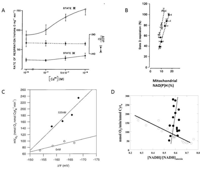
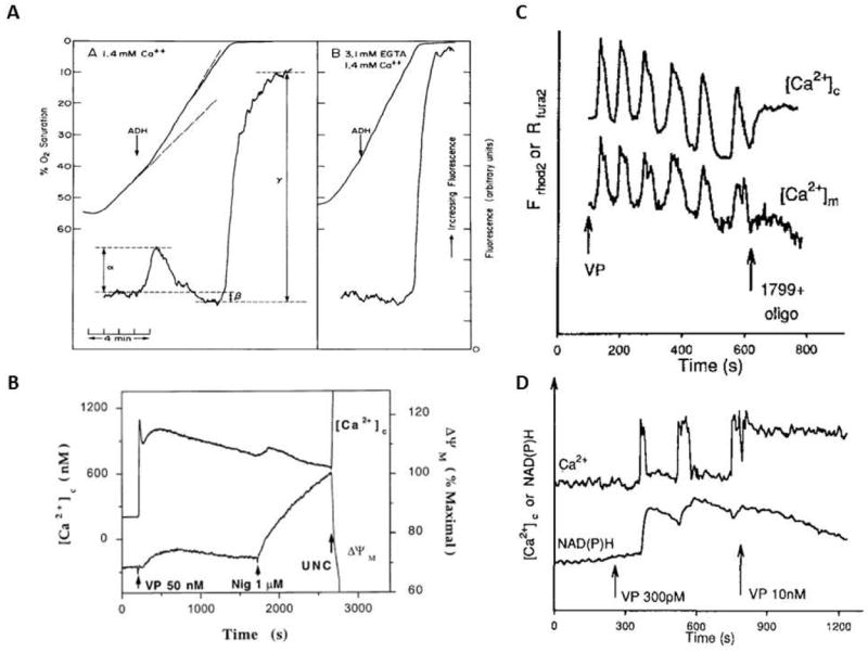
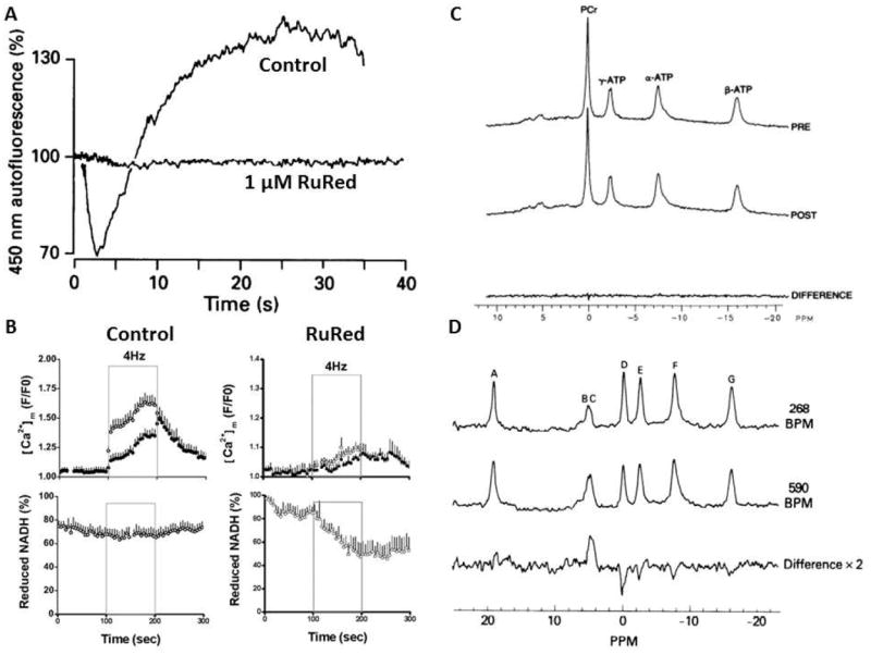
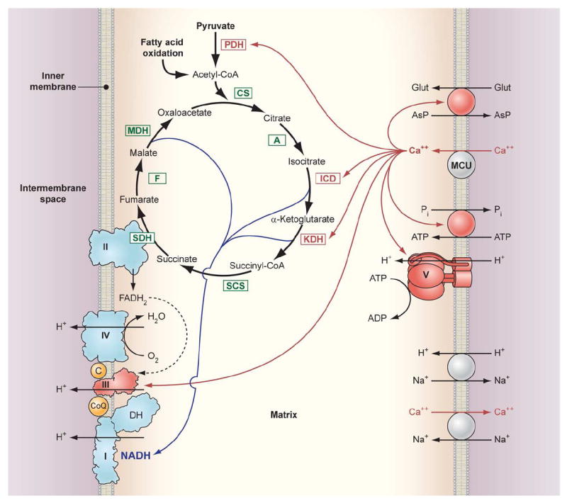
Calcium is an important signaling molecule involved in the regulation of many cellular functions. The large free energy in the Ca(2+) ion membrane gradients makes Ca(2+) signaling inherently sensitive to the available cellular free energy, primarily in the form of ATP. In addition, Ca(2+) regulates many cellular ATP-consuming reactions such as muscle contraction, exocytosis, biosynthesis, and neuronal signaling. Thus, Ca(2+) becomes a logical candidate as a signaling molecule for modulating ATP hydrolysis and synthesis during changes in numerous forms of cellular work. Mitochondria are the primary source of aerobic energy production in mammalian cells and also maintain a large Ca(2+) gradient across their inner membrane, providing a signaling potential for this molecule. The demonstrated link between cytosolic and mitochondrial Ca(2+) concentrations, identification of transport mechanisms, and the proximity of mitochondria to Ca(2+) release sites further supports the notion that Ca(2+) can be an important signaling molecule in the energy metabolism interplay of the cytosol with the mitochondria. Here we review sites within the mitochondria where Ca(2+) plays a role in the regulation of ATP generation and potentially contributes to the orchestration of cellular metabolic homeostasis. Early work on isolated enzymes pointed to several matrix dehydrogenases that are stimulated by Ca(2+), which were confirmed in the intact mitochondrion as well as cellular and in vivo systems. However, studies in these intact systems suggested a more expansive influence of Ca(2+) on mitochondrial energy conversion. Numerous noninvasive approaches monitoring NADH, mitochondrial membrane potential, oxygen consumption, and workloads suggest significant effects of Ca(2+) on other elements of NADH generation as well as downstream elements of oxidative phosphorylation, including the F(1)F(O)-ATPase and the cytochrome chain. These other potential elements of Ca(2+) modification of mitochondrial energy conversion will be the focus of this review. Though most specific molecular mechanisms have yet to be elucidated, it is clear that Ca(2+) provides a balanced activation of mitochondrial energy metabolism that exceeds the alteration of dehydrogenases alone.


Figures

Figure 1
Figure 1
A) Effect of extra-mitochondrial Ca2+ on the rate of respiration in isolated rat liver mitochondria respiring on 5 mM succinate. A 5 minute pre-incubation with Ca2+ was used before driving State 3 respiration with 250 μM ADP(74). Reproduced with permission. B) Effect of extramitochondrial Ca2+ on the relationship between [NAD(P)H] and respiration in isolated liver mitochondria. All experiments were conducted with 5 mM malate. Solid points represent the control study with effectively no Ca2+ and mitochondrial [NAD(P)] increased with increasing glutamate (0.25, 0.5, 1, 2, 5 and 10 mM). The open circles are with fixed substrates 1 mM glutamate and 5 mM malate with the extramitochondrial Ca2+ varied from 5 nM (initial value), 66, 130, 225 and 400 nM (adapted from(83) reproduced with permission) C) Mitochondrial membrane potential versus oxygen consumption of State-3 respiration with variable substrates in the presence and absence of Ca2+in porcine heart mitochondria. In all experiments only the glutamate /malate total concentration was varied from 0.5-5.0 mM to generate a difference in driving force from the citric acid cycle. The open symbols are in the nominal absence of Ca2+. The filled symbols are in the presence of the previously determined optimal Ca2+ concentration of 535 nM. Data from (87), reproduced with permission. D) Relationship between [NADH] and oxygen consumption with apyrase or a sarcoplasmic reticulum(SR)-mitochondria reconstitution system Open symbols are data collected by varying [Apyrase]. Increasing [Apyrase] increased respiration but decreased [NADH]. Closed Signals are fixed [SR] and [mitochondria] (ratio of SR to mitochondria 0.5) with varying [Ca2+] over low physiological levels of 0 to 492 nM (calculated free concentration). Increasing [Ca2+] increased respiration over 5 fold with a slight increase in [NADH]. Lines are the linear regression of the data points. Data from (92), reproduced with permission.
Figure 2
Figure 2
A) Effect of ADH on NADH and oxygen consumption of isolated rat hepatocytes. In both panels 100 nM ADH was added to the chambers at the arrow in the absence (A) and presence (B) of 3.1 mM EGTA. Data from (110), reproduced with permission. B) Measurements of Ca2+c and ΔΨ in cultured hepatocytes with vasopressin (50 nM). [Ca2+]c was measured using fura-2 and ΔΨ was monitored with TMREE. Data from (111), reproduced with permission. C) Temporal correlation of Ca2+c and Ca2+m during hormonal stimulation of hepatocytes. [Ca2+]m was monitored in hepatocytes loaded with dihydro-Rhod 2-AM, and [Ca2+]c was measured in cells loaded with Fura 2-AM. Data from (114), reproduced with permission. D) 4. Relationship between the frequency of Ca2+c and NAD(P)Hm fluorescence. NAD(P)H and [Ca2+]c were measured simultaneously during the addition of vasopressin (VP). Data from (114), reproduced with permission
Figure 3
Figure 3
A) Effect of patch clamp depolarization (from -70 mV holding potential to 0 mV) of freshly isolated neurons NAD(P)H fluorescence in the absence and presence of RuRed. RuRed did not block the cellular membrane Ca2+ current associated with the depolarization but blocked the NAD(P)H fluorescence response. Data adapted from(117), reproduced with permission. B) Effect of 4 Hz pacing on isolated cardiac myocytes NADH fluorescence and Ca2+m in the absence (control) and presence RuRed. The pacing of the myocytes occurred at the times indicated. Data from (118) reproduced with permission. C) Effect of pacing work on the 31P NMR spectrum of the intact canine heart. Top spectrum represents the control spectrum while the post spectrum represents the average 31P NMR spectrum during pacing the heart to maximum pacing induced coronary blood flow and oxygen consumption. The bottom trace is the difference of the control and maximum pacing protocol. Data from(141), reproduced with permission. D) Effect of RuRed on the 31P NMR spectrum of the perfused rat heart undergoing an increase in pacing rate. The top spectrum is during the control pacing rate, middle spectrum was collected during the increase in pacing rate to 580 beats/min, bottom spectrum represents the difference between these two conditions with a large increase in Pi and decrease in CrP. The peak assignments are A: external reference, B-C: Pi, D: CrP, E: γP-ATP, F: αP-ATP/NAD and G: βP-ATP. Data from (146), reproduced with permission.
Figure 4
Figure 4
Schematic diagram of the interaction of matrix Ca2+ with processes involved in oxidative phosphorylation. Enzyme abbreviations: PDH: Pyruvate dehydrogenase, CS: Citrate synthase, A: Aconitase, ICD: Isocitrate dehydrogenase, KDH: α ketoglutarate dehydrogenase, SCS: Succinyl CoA synthetase, SDH: Succinate dehydrogenase (also Complex II), F: Fumarase, MDH: Malate dehydrogenase, MCU: Mitochondria calcium uniporter. The Complexes of oxidative phosphorylation are labeled as roman numerals from I-V. The DH on Complex 1 refers to the intrinsic and possibly associated NADH dehydrogenase activity. The red arrows from Ca2+ to the different interaction sites imply either a direct or indirection modulation of the transport or enzymatic activity.

References

    1. Hasselbach W, Oetliker H. Energetics and electrogenicity of the sarcoplasmic reticulum calcium pump. Annu Rev Physiol. 1983;45:325–339. - PubMed
    1. Chen W, London R, Murphy E, Steenbergen C. Regulation of the Ca2+ gradient across the sarcoplasmic reticulum in perfused rabbit heart. A 19F nuclear magnetic resonance study. Circ Res. 1998;83:898–907. - PubMed
    1. Tian R, Halow JM, Meyer M, Dillmann WH, Figueredo VM, Ingwall JS, Camacho SA. Thermodynamic limitation for Ca2+ handling contributes to decreased contractile reserve in rat hearts. Am J Physiol. 1998;275:H2064–H2071. - PubMed
    1. Hill AV. A challange to biochemists. Biochim Biophys Acta. 1950;4:4–11. - PubMed
    1. Devin A, Rigoulet M. Mechanisms of mitochondrial response to variations in energy demand in eukaryotic cells. Am J Physiol Cell Physiol. 2007;292:C52–C58. - PubMed