. 2012 Mar 25:13:14.
doi: 10.1186/1471-2369-13-14.

Acatalasemic mice are mildly susceptible to adriamycin nephropathy and exhibit increased albuminuria and glomerulosclerosis

Affiliations

Acatalasemic mice are mildly susceptible to adriamycin nephropathy and exhibit increased albuminuria and glomerulosclerosis

Keiichi Takiue et al. BMC Nephrol. .

Abstract

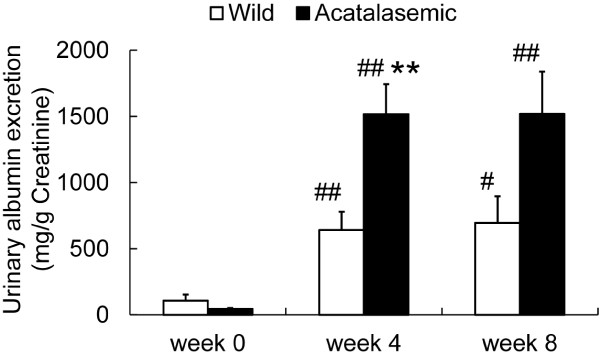
Background: Catalase is an important antioxidant enzyme that regulates the level of intracellular hydrogen peroxide and hydroxyl radicals. The effects of catalase deficiency on albuminuria and progressive glomerulosclerosis have not yet been fully elucidated. The adriamycin (ADR) nephropathy model is considered to be an experimental model of focal segmental glomerulosclerosis. A functional catalase deficiency was hypothesized to exacerbate albuminuria and the progression of glomerulosclerosis in this model.

Methods: ADR was intravenously administered to both homozygous acatalasemic mutant mice (C3H/AnLCs(b)Cs(b)) and control wild-type mice (C3H/AnLCs(a)Cs(a)). The functional and morphological alterations of the kidneys, including albuminuria, renal function, podocytic, glomerular and tubulointerstitial injuries, and the activities of catalase were then compared between the two groups up to 8 weeks after disease induction. Moreover, the presence of a mutation of the toll-like receptor 4 (tlr4) gene, which was previously reported in the C3H/HeJ strain, was investigated in both groups.

Results: The ADR-treated mice developed significant albuminuria and glomerulosclerosis, and the degree of these conditions in the ADR-treated acatalasemic mice was higher than that in the wild-type mice. ADR induced progressive renal fibrosis, renal atrophy and lipid peroxide accumulation only in the acatalasemic mice. In addition, the level of catalase activity was significantly lower in the kidneys of the acatalasemic mice than in the wild-type mice during the experimental period. The catalase activity increased after ADR injection in wild-type mice, but the acatalasemic mice did not have the ability to increase their catalase activity under oxidative stress. The C3H/AnL strain was found to be negative for the tlr4 gene mutation.

Conclusions: These data indicate that catalase deficiency plays an important role in the progression of renal injury in the ADR nephropathy model.


Figures

Figure 1
Figure 1
The changes in urinary albumin excretion (UAE) in wild-type (open bars) and acatalasemic mice (closed bars). The UAE significantly increased in both groups at 4 and 8 weeks after Adriamycin (ADR) administration. The elevation of UAE in acatalasemic mice was higher at 4 weeks after ADR administration compared to that in wild-type mice. Each column consists of the means ± SE. N = 9 to 39 animals/group. **: P < 0.01 vs. wild-type ADR mice at the same time point. #: p < 0.05 vs. week 0 in the same group. ##: p < 0.01 vs. week 0 in the same group.
Figure 2
Figure 2
The renal histology in wild-type and acatalasemic mice. Light micrographs of a glomerulus of wild-type (A, C and E) and acatalasemic (B, D and F) glomeruli at 0, 4 and 8 weeks after adriamycin (ADR) administration are shown. The week 0 glomeruli were microscopically normal. Note that the segmental sclerosis is significant in both acatalasemic and wild-type glomeruli at the later time points. The glomerular area (G) and sclerosis index (H) of wild-type (open bars) or acatalasemic (closed bars) mice are also shown. A through F: periodic acid-Schiff stain. Scale bars: 20 μm (A through F). G and H: Each column shows the means ± SE. N = 6 to 15 animals/group. *: P < 0.05 vs. wild-type ADR mice at the same time point. #: p < 0.05 vs. week 0 in the same group. ##: p < 0.01 vs. week 0 in the same group.
Figure 3
Figure 3
Light micrographs of the wild-type (A, C and E) and acatalasemic (B, D and F) kidney cortex at 0, 4 and 8 weeks after adriamycin (ADR) administration are shown. Note that interstitial fibrosis significantly developed in the kidneys of the acatalasemic mice. The fibrosis scores (G) and cast scores (H) of wild-type (open bars) and acatalasemic (closed bars) mice are also shown. A through F: Masson's trichrome stain. Scale bars: 200 μm (A through F). G and H: Each column includes the means ± SE. N = 6 to 15 animals/group. #: p < 0.05 vs. week 0 in the same group. ##: p < 0.01 vs. week 0 in the same group.
Figure 4
Figure 4
Immunohistochemical staining of 4-hydroxy-2-nonenal (4-HNE). Wild-type (A, C and E) and acatalasemic (B, D and F) cortex samples at 0, 4 and 8 weeks after adriamycin (ADR) administration are shown. The 4-HNE positive areas (G) of wild-type (open bars) or acatalasemic (closed bars) mice are also shown. Scale bars: 50 μm (A through F). G: Each column shows the means ± SE. N = 6 to 8 animals/group. **: P < 0.01 vs. wild-type ADR mice at the same time point. ##: p < 0.01 vs. week 0 in the same group.
Figure 5
Figure 5
The renal catalase activities in wild-type (open bars) and acatalasemic mice (closed bars). Each column consists of the means ± SE. N = 5 to 10 animals/group. *: P < 0.05 vs. wild-type adriamycin (ADR) mice at the same time point. **: P < 0.01 vs. wild-type ADR mice at the same time point. ##: p < 0.01 vs. week 0 in the same group.

References

    1. Chance B, Sies H, Boveris A. Hydroperoxide metabolism in mammalian organs. Physiol Rev. 1979;59(3):527–605. - PubMed
    1. Zamocky M, Furtmuller PG, Obinger C. Evolution of catalases from bacteria to humans. Antioxid Redox Signal. 2008;10(9):1527–1548. doi: 10.1089/ars.2008.2046. - DOI - PMC - PubMed
    1. Takahara S. Progressive oral gangrene probably due to lack of catalase in the blood (acatalasaemia); report of nine cases. Lancet. 1952;2(6745):1101–1104. - PubMed
    1. Ogata M. Acatalasemia. Hum Genet. 1991;86(4):331–340. - PubMed
    1. Ogata M, Wang DH, Ogino K. Mammalian acatalasemia: the perspectives of bioinformatics and genetic toxicology. Acta Med Okayama. 2008;62(6):345–361. - PubMed

Publication types