Review
. 2012:35:203-25.
doi: 10.1146/annurev-neuro-062111-150444. Epub 2012 Mar 20.

Mechanisms of gamma oscillations

Affiliations
Review

Mechanisms of gamma oscillations

György Buzsáki et al. Annu Rev Neurosci. 2012.

Abstract

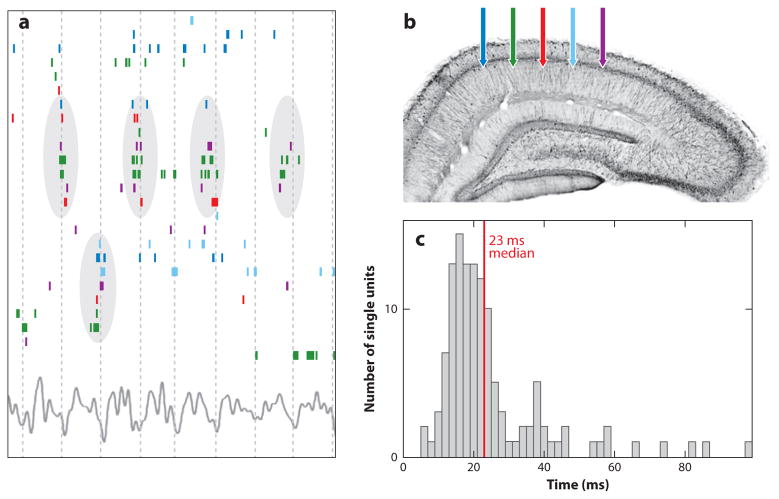
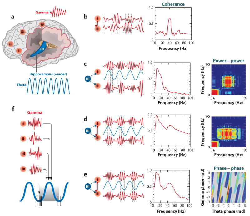
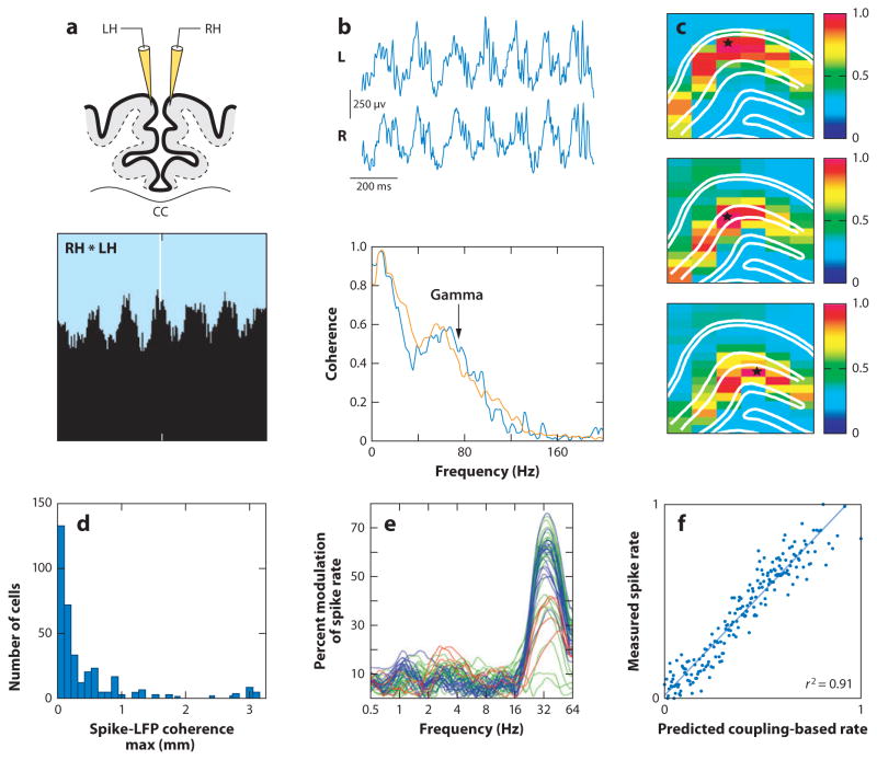
Gamma rhythms are commonly observed in many brain regions during both waking and sleep states, yet their functions and mechanisms remain a matter of debate. Here we review the cellular and synaptic mechanisms underlying gamma oscillations and outline empirical questions and controversial conceptual issues. Our main points are as follows: First, gamma-band rhythmogenesis is inextricably tied to perisomatic inhibition. Second, gamma oscillations are short-lived and typically emerge from the coordinated interaction of excitation and inhibition, which can be detected as local field potentials. Third, gamma rhythm typically concurs with irregular firing of single neurons, and the network frequency of gamma oscillations varies extensively depending on the underlying mechanism. To document gamma oscillations, efforts should be made to distinguish them from mere increases of gamma-band power and/or increased spiking activity. Fourth, the magnitude of gamma oscillation is modulated by slower rhythms. Such cross-frequency coupling may serve to couple active patches of cortical circuits. Because of their ubiquitous nature and strong correlation with the "operational modes" of local circuits, gamma oscillations continue to provide important clues about neuronal population dynamics in health and disease.


Figures

Figure 1
Figure 1
Dynamical cell assemblies are organized in gamma waves. (a) Raster plot of a subset of hippocampal pyramidal cells that were active during a 1-s period of spatial exploration on an open field out of a larger set of simultaneously recorded neurons, ordered by stochastic search over all possible orderings to highlight the temporal relationship between anatomically distributed neurons. Color-coded ticks (spikes) refer to recording locations shown in panel b. Vertical lines indicate troughs of theta waves (bottom trace). Cell-assembly organization is visible, with repeatedly synchronous firing of some subpopulations (circled). (c) Spike timing is predictable from peer activity. Distribution of timescales at which peer activity optimally improved spike-time prediction of a given cell, shown for all cells. The median optimal timescale is 23 ms (red line). Based on Harris et al. (2003).
Figure 2
Figure 2
I-I and E-I models of gamma oscillations. (a) Clock-like rhythm of coupled oscillators in an interneuronal (I-I) population. (Upper panel) Single interneurons fire spikes periodically at ~40 Hz. Mutual inhibition via GABAA receptors quickly brings them to zero-phase synchrony; (lower panel) two example neurons. Adapted from Wang & Buzsáki (1996). (b,c) Sparsely synchronous oscillations in a neural circuit where single neuronal spiking is stochastic. Adapted from Geisler et al. (2005). (b) Interneuronal population in noise-dominated regime typically exhibits gamma power in the higher frequency range, in contrast to (a) the clock-like rhythmic case. (c) Reciprocally connected E-I network where pyramidal cells send fast excitation via AMPA receptors to interneurons, which in turn provide inhibition via GABAA receptors, leading to coherent oscillations in the gamma-frequency range.
Figure 3
Figure 3
A critical role of parvalbumin (PV) basket cells in gamma oscillations. (a) Local field potential (LFP) recording from the CA1 pyramidal layer (top) and dentate hilus (bottom) and unit recording from a fast-spiking putative interneuron in the hilus (middle trace). Note the trains of spikes at gamma frequency, repeating periodically at theta frequency. (b) Power spectrum of the unit shown in panel a. Note the peak at theta and a broader peak at 50–80 Hz (gamma). (c) Spike-triggered average of the LFP in the hilus. Note the prominent phase locking of the interneuron to gamma wave phase and the cross-frequency coupling between gamma and theta waves. (ac) Recordings from a behaving rat. (d) Camera-lucida reconstruction of the axon arbor of an immunocytochemically identified CA1 basket cell in vivo. The axon arbor outlines the CA1 pyramidal layer, showing (circles) putative contacts with other PV-positive neurons, (inset) averages of the intracellularly recorded Vm (membrane potential) and the LFP, (triangle) peak of the mean preferred discharge of the surrounding pyramidal cells, and (arrow) peak of the mean preferred discharge of the basket cell. Note the short delay between the spikes of pyramidal cells and the basket neuron. Current source density (CSD) map is superimposed on the pyramidal layer. Arrow points to current source of gamma wave (red). (e) Continuous display (110) of integrated and rectified gamma activity of the LFP and the fast intracellularly recorded Vm fluctuation (20–80 Hz; after digital removal of spikes) in a CA1 pyramidal neuron. Vm was biased by the intracellular current injection: (dashed line) resting membrane potential. Note the increase of the intracellular Vm gamma during both depolarization (inset) and hyperpolarization as well as the smallest Vm gamma power at resting membrane potential (asterisks) against the steady background of LFP gamma power. (d, e) In vivo recordings under urethane anesthesia. (f) Excitatory (E) and inhibitory (I) postsynaptic currents (PSCs) in a pyramidal cell, triggered by LFP gamma (top) and the spike timing of a pyramidal cell (P) and a basket interneuron (B) during carbachol-induced gamma oscillation in a hippocampal slice in vitro. Note that maximum discharge of the basket cell precedes the hyperpolarization of the pyramidal cell. (g) Intracellular recordings in a ferret prefrontal pyramidal cell in vivo illustrating the large amplitude, inhibition-dominated barrages recorded at 0 mV (brown) and smaller amplitude, excitation-dominated, synaptic barrages recorded at −80 mV (tan) for two representative UP states. Membrane potentials are expanded further (inset). EPSP, excitatory postsynaptic potential; IPSP, inhibitory postsynaptic potential. Reproduced with permission from (ac) Buzsáki et al. (1983), (de) after Penttonen et al. (1998), (f) after from Mann et al. (2005), and (g) after Hasenstaub et al. (2005).
Figure 4
Figure 4
“Synthetic” gamma rhythm in vivo. (a) Local field potential (LFP) recordings in anesthetized mouse, expressing ChR2 selectively in either parvalbumin (PV) neurons (ChR2-PV-Cre) or pyramidal cells (ChR2-αCamKII-Cre). Stimulation at 8 Hz evoked rhythmic activity in the αCamKII-Cre but not the PV-Cre mouse. Conversely, stimulation at 40 Hz induced gamma oscillation in the PV-Cre but not in αCamKII-Cre mouse. (b) Mean LFP power ratio measured in multiple frequency bands in response to rhythmic light activation of ChR2-PV-Cre expressing neurons (blue) or ChR2-αCamKII-Cre expressing neurons (purple) at various frequencies. Reprinted from Cardin et al. (2009).
Figure 5
Figure 5
Oscillatory coupling mechanisms. (a) Schematic view of the human brain showing hot spots of transient gamma oscillations (i–iv) and theta oscillation in the hippocampus (HI); entorhinal cortex (EC). Oscillators of the same and different kind (e.g., theta, gamma) can influence each other in the same and different structures, thereby modulating the phase, amplitude, or both. (b) Phase-phase coupling of gamma oscillations between two areas. Synthetic data used for illustration purposes. Coherence spectrum (or other, more specific, phase-specific measures) between the two signals can determine the strength of phase coupling. (c) Cross-frequency phase-amplitude coupling. Although phase coupling between gamma waves is absent, the envelope of gamma waves at the two cortical sites is modulated by the common theta rhythm. This can be revealed by the power-power correlation (comodugram; right). (d) Gamma phase-phase coupling between two cortical sites, whose powers are modulated by the common theta rhythm. Both gamma coherence and gamma power-power coupling are high. (e) Cross-frequency phase-phase coupling. Phases of theta and gamma oscillations are correlated, as shown by the phase-phase plot of the two frequencies. (f) Hippocampal theta oscillation can modulate gamma power by its duty cycle at multiple neocortical areas so that the results of the local computations are returned to the hippocampus during the accrual (“readiness”) phase of the oscillation. a and f, after Buzsáki (2010); be, after Belluscio et al. (2012).
Figure 6
Figure 6
Long-range synchrony of gamma oscillations. (a) Neurons sharing receptive fields in left (LH) and right (RH) primary visual cortex of the anesthetized cat fire coherently with zero time lag at gamma frequency. (b) Local field potential (LFP) traces from the left (L) and right (R) hippocampal CA1 pyramidal layer of the mouse during running and coherence spectra between the traces during running (orange) and REM sleep (blue). (c) LFP coherence map of gamma (30–90 Hz) in the rat hippocampus during running. Coherence was calculated between the reference site (star) and the remaining 96 recording sites. Note the high coherence values within the same layers (outlined by white lines) and rapid decrease of coherence across layers. (d) Distribution of distances between the unit and LFP recording sites with maximum spike-LFP coherence in the gamma band. Note that, in a fraction of cases, maximum coherence is stronger at large distances between the recorded unit and the LFP. (e) Spike-LFP coherence in the human motor cortex. The probability of spiking correlates with frequency-specific LFP phase of the ipsilateral (blue) and contralateral (green) motor area and contralateral dorsal premotor area (red). (f) The phase-coupling-based spike rate (generated from the preferred LFP–LFP phase-coupling pattern) predicts the measured spike rate. Panels reproduced after (a) Engel et al. (1991), (b) Buzsáki et al. (2003), (c) Montgomery & Buzsáki (2007), (d) Sirota et al. (2008), and (e,f) Canolty et al. (2010).
Figure 7
Figure 7
Coupling of gamma oscillators by long-range interneurons. (a) Oscillations in a network with locally connected interneurons. The network is essentially asynchronous. (Upper panel) Spike raster of 4000 neurons; (lower panel) the population firing rate. (b) Oscillations in a network with local interneurons (B) and long-range interneurons (LR; power-law connectivity). Note clear oscillatory rhythm. (c) Cross-section of the axon of a long-range CA1 GABAergic interneuron projecting toward the subiculum/entorhinal cortex. In comparison, neighboring axons of pyramidal cells are also shown (d). Reproduced from Buzsáki et al. (2004) (a,b) and from Jinno et al. (2006) (b,c).
Figure 8
Figure 8
Multiple gamma sub-bands. Wavelet power between 30 and 150 Hz as a function of waveform-based theta cycle phases. Note the different theta-phase preference of mid-frequency (M) (gammaM, 50–90 Hz, near theta peak) and slow (S) (gammaS, 30–50 Hz on the descending phase of theta) gamma oscillations. Note also the dominance of fast (F) (gammaF, or epsilon band, 90–150 Hz) at the trough of theta. After Belluscio et al. (2012).

References

LITERATURE CITED

    1. Adrian E. Olfactory reactions in the brain of the hedgehog. J Physiol. 1942;100:459–73. - PMC - PubMed
    1. Ardid S, Wang X-J, Gomez-Cabrero D, Compte A. Reconciling coherent oscillation with modulation of irregular spiking activity in selective attention: gamma-range synchronization between sensory and executive cortical areas. J Neurosci. 2010;30(8):2856–70. - PMC - PubMed
    1. Axmacher N, Henseler MM, Jensen O, Weinreich I, Elger CE, Fell J. Cross-frequency coupling supports multi-item working memory in the human hippocampus. Proc Natl Acad Sci USA. 2010;107:3228–33. - PMC - PubMed
    1. Bartos M, Vida I, Jonas P. Synaptic mechanisms of synchronized gamma oscillations in inhibitory interneuron networks. Nat Rev Neurosci. 2007;8:45–56. - PubMed
    1. Belluscio MA, Mizuseki K, Schmidt R, Kempter R, Buzsáki G. Cross-frequency phase-phase coupling between theta and gamma oscillations in the hippocampus. J Neurosci. 2012;32(2):423–35. - PMC - PubMed

RELATED RESOURCES

    1. Bartos M, Vida I, Jonas P. Synaptic mechanisms of synchronized gamma oscillations in inhibitory interneuron networks. Nat Rev Neurosci. 2007;8:45–56. - PubMed
    1. Engel A, Fries P, Singer W. Dynamic predictions: oscillations and synchrony in top-down processing. Nat Rev Neurosci. 2001;2:704–16. - PubMed
    1. György Buzsáki’s Web page. http://www.med.nyu.edu/buzsakilab/
    1. Uhlhaas P, Singer W. Neural synchrony in brain disorders: relevance for cognitive dysfunctions and pathophysiology. Neuron. 2006;52:155–68. - PubMed
    1. Varela F, Lachaux J, Rodriguez E, Martinerie J. The brainweb: phase synchronization and large-scale integration. Nat Rev Neurosci. 2001;2:229–39. - PubMed

Publication types