Review
. 2012:81:767-93.
doi: 10.1146/annurev-biochem-072909-095555. Epub 2012 Mar 23.

Endoplasmic reticulum stress and type 2 diabetes

Affiliations
Review

Endoplasmic reticulum stress and type 2 diabetes

Sung Hoon Back et al. Annu Rev Biochem. 2012.

Abstract

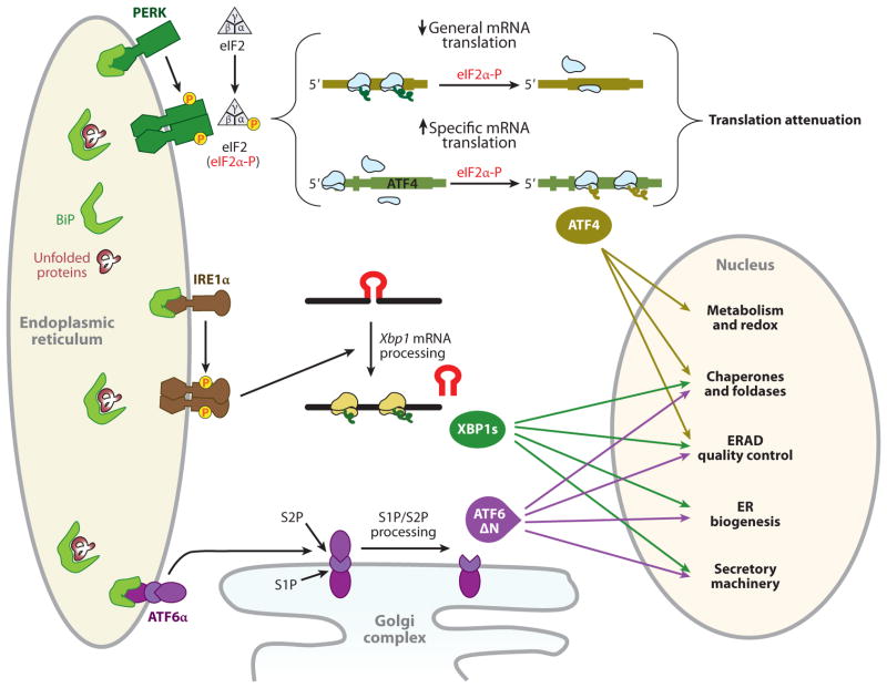
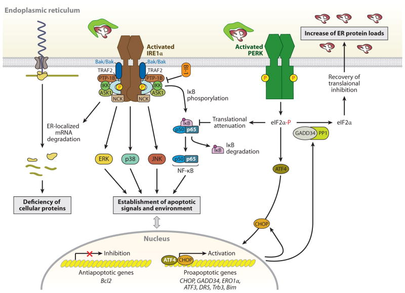
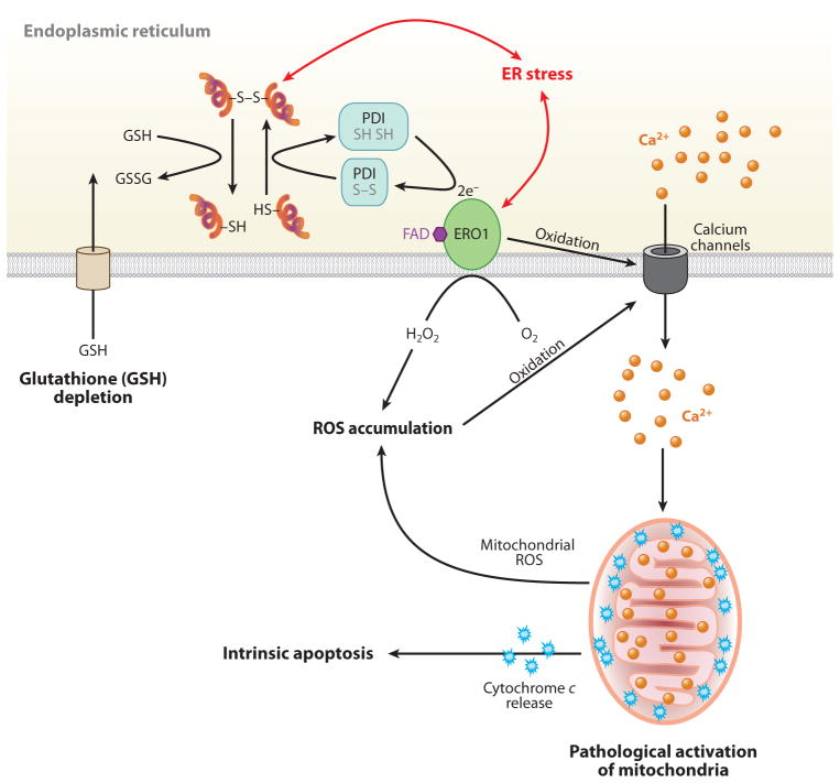
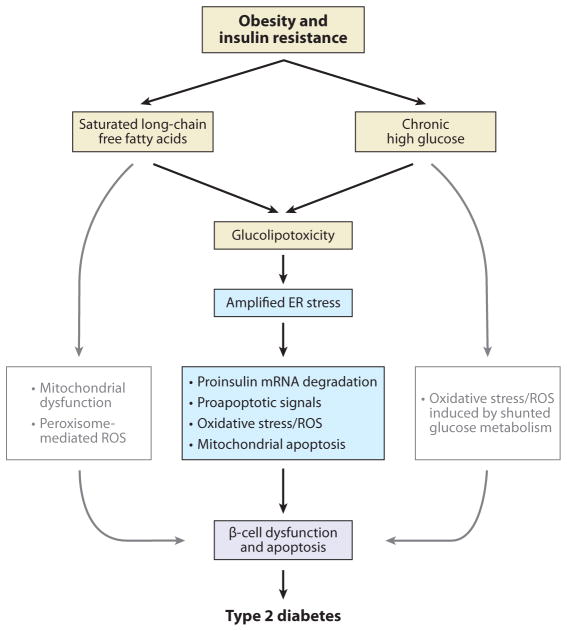
Given the functional importance of the endoplasmic reticulum (ER), an organelle that performs folding, modification, and trafficking of secretory and membrane proteins to the Golgi compartment, the maintenance of ER homeostasis in insulin-secreting β-cells is very important. When ER homeostasis is disrupted, the ER generates adaptive signaling pathways, called the unfolded protein response (UPR), to maintain homeostasis of this organelle. However, if homeostasis fails to be restored, the ER initiates death signaling pathways. New observations suggest that both chronic hyperglycemia and hyperlipidemia, known as important causative factors of type 2 diabetes (T2D), disrupt ER homeostasis to induce unresolvable UPR activation and β-cell death. This review examines how the UPR pathways, induced by high glucose and free fatty acids (FFAs), interact to disrupt ER function and cause β-cell dysfunction and death.


Figures

Figure 1
Figure 1
The adaptive unfolded protein response (UPR). Activation of three UPR pathways initiates the adaptive endoplasmic reticulum (ER) stress response. During activation of the UPR in mammals, BiP (immunoglobulin heavy chain binding protein, also known as GRP78) is sequestered through binding to unfolded or misfolded polypeptide chains, thereby leading to BiP release from the ER stress sensors for their activation. Unconventional cytoplasmic splicing, mediated by IRE1α, removes a 26-nucleotide intron from unspliced X-box-binding protein 1 (Xbp1) mRNA (encoding 267 amino acids) to produce a translational frameshift, yielding a fusion protein encoded from two evolutionarily conserved open reading frames (16). The fusion protein, XBP1s, acts as a potent transcription factor for expression of UPR target genes involved in protein folding and export from the ER, export and degradation of misfolded proteins, and lipid biosynthesis, to resolve ER stress (16). Upon accumulation of unfolded protein in the ER lumen, oligomerization of the PKR-like ER kinase (PERK) in ER membranes induces its autophosphorylation and kinase domain activation (141, 142). Activated PERK phosphorylates serine 51 on the α-subunit of heterotrimeric eIF2 (143). When eukaryotic translation initiation factor 2α (eIF2α) is phosphorylated, the eIF2 complex shows increased affinity for its guanine nucleotide exchange factor eIF2B and sequesters all available eIF2B. Because the cellular level of eIF2B is 10- to 20-fold lower than the level of eIF2, very small changes in eIF2α phosphorylation can dramatically change the rate of translation initiation (144). Inhibition of general mRNA translation by the phosphorylation of eIF2α reduces accumulation of misfolded protein in the ER lumen (22), thereby protecting the cell from diverse stimuli that perturb the ER homeostasis. In contrast to inhibition of general mRNA translation, the PERK/eIF2α pathway stimulates the translation of several specific mRNAs containing multiple 53-upstream open reading frames, such as Atf4 and Atf5, Chop, Gadd34, and the cationic amino acid transporter 1 (Cat-1, an Na+-independent transporter of L-arginine and L-lysine) (28, 145). Among them, ATF4 activates transcription of the adaptive genes that encode functions in ER protein folding, endoplasmic reticulum–associated degradation (ERAD), amino acid biosynthesis and transportation, and the antioxidative stress response (24). Under ER stress, ATF6α and ATF6βare released from BiP and translocate to the Golgi complex, where they are cleaved by Golgi-resident proteases, first by S1P (site 1 protease) and then in the intramembrane region by S2P (site 2 protease), to release the N-terminal basic leucine zipper protein (bZIP) transcription factor domain (16). The bZIP domain of ATF6α then translocates into the nucleus, where it activates the transcription of genes encoding ER-localized molecular chaperones and folding enzymes, ERAD, protein secretion machineries, and ER biogenesis (146), in some cases in cooperation with XBP1s (42).
Figure 2
Figure 2
IRE1α- and PERK-mediated cell death pathways. During endoplasmic reticulum (ER) stress, inositol-requiring protein 1α (IRE1α) forms a hetero-oligomeric complex with TNF receptor-associated factor 2 (TRAF2) (76) and apoptosis signal-regulating kinase 1 (ASK1) (77) and then recruits the protein kinase JNK, leading to the activation of JNK (76). The IRE1α-TRAF2 complex recruits IκB kinase (IKK), which phosphorylates inhibitor of κB (IκB), leading to the degradation of IκB and the nuclear translocation of nuclear factor κ-light-chain-enhancer of activated B cells (NF-κB) (147). IRE1α also modulates the activation of other “alarm genes,” such as p38 and ERK (80), possibly by the binding of the Src homology (SH) 2/3 -containing adaptor proteins Nck and TRAF2, respectively. Furthermore, several proapoptotic (i.e., BAX/BAK, AIP1, and maybe PTP-1B) or antiapoptotic proteins (i.e., BI-1) interact with IRE1α, regulating its activation state (–84). Thus, the formation of a macromolecular signaling complex of IRE1α with several proapoptotic proteins can generate apoptotic signals and establish an apoptotic environment. In addition, the endoribonuclease activity of IRE1α, aside from specific cleavage of Xbp1 mRNA, degrades ER-targeted mRNAs that can decrease cellular functions, such as proinsulin synthesis in β-cells. In contrast to the adaptive response by the PERK-phosphorylated eIF2α-ATF4 pathway, this pathway also contributes to stress-induced cell death by ATF4-mediated induction of proapoptotic genes, including CHOP, ATF3, and GADD34 (16). The induced transcription factor CHOP contributes to increased expression of the proapoptotic factors, such as death receptor 5 (DR5) (148), tribbles-related protein 3 (Trb3) (149), and binding to microtubule (Bim) (150), and it can suppress B cell lymphoma 2 (Bcl2) expression. Bim is also activated through protein phosphatase 2A-mediated dephosphorylation, which prevents its ubiquitination and proteasomal degradation (150). PERK-mediated translational attenuation upregulates NF-κB-dependent transcription because IκB has a shorter half-life than NF-κB, so NF-κB is released to translocate to the nucleus (151). Recovery from translational repression is mediated by eIF2α dephosphorylation by the two regulatory subunits of protein phosphatase 1 (PP1), GADD34 and CReP (constitutive repressor of eIF2α phosphorylation) (16). GADD34 is induced transcriptionally during ER stress by ATF4, whereas CreP is a constitutive activator of PP1. The premature dephosphorylation of eIF2α by the GADD34-PP1 complex restores translation of general mRNAs, which may be detrimental if the ER protein-folding defect is not resolved.
Figure 3
Figure 3
The role of calcium and reactive oxygen species (ROS) in endoplasmic reticulum (ER) stress-mediated cell death. In the ER lumen, oxidative protein folding is catalyzed by protein disulfide isomerase (PDI) and ER oxidoreductase (ERO1). In this reaction, an oxidant flavin adenine dinucleotide (FAD)-bound ERO1 oxidizes PDI, which subsequently oxidizes folding proteins directly. FAD-bound ERO1 then passes two electrons to molecular oxygen, resulting in the production of hydrogen peroxide (73). During unfolded protein response activation, CHOP-mediated induction of ERO1α hyperoxidizes the ER lumen and causes oxidation-induced activation of the ER Ca2+ release channel inositol 1,4,5-trisphosphate receptor (152), causing a large and transient release of Ca2+ from the ER. Increased cytosolic Ca2+ is taken up into the mitochondrial matrix, and this stimulates mitochondrial ROS production through disruption of mitochondrial electron transport (153). High levels of ROS generated from mitochondria, in turn, further increase Ca2+ release from the ER. The increase in mitochondrial Ca2+ eventually dissociates cytochrome c from the inner membrane cardiolipin, which triggers permeability transition pore opening and cytochrome c release across the outer membrane. Now the vicious cycle of ER calcium release and mitochondrial ROS production activates cytochrome c-mediated apoptosis. In addition, ER stress may cause consumption of excessive cellular glutathione (GSH) because reduced GSH may also assist in reducing nonnative disulfide bonds in misfolded proteins, resulting in the production of oxidized glutathione (GSSG).
Figure 4
Figure 4
Apoptotic unfolded protein response (UPR) pathways induced by free fatty acids (FFAs) and chronically high glucose in β-cells. In contrast to unsaturated FFAs, saturated FFAs serve as poor substrates for mitochondrial fatty acid oxidation and de novo triglyceride synthesis. However, saturated FFAs serve as intermediates in ceramide biosynthesis. The saturated FFAs activate UPR pathways (primarily PKR-like ER kinase, PERK) by perturbation of ER Ca2+ mobilization through inhibition of sarco/endoplasmic reticulum Ca2+-ATPase (SERCA), or activation of inositol-1,4,5-trisphosphate (IP3) receptors, and/or direct impairment of endoplasmic reticulum (ER) homeostasis. In addition, chronically high glucose increases biosynthesis of proinsulin and islet amyloid polypeptide in β-cells, which increases accumulation of misfolded proteins [insulin and islet amyloid polypeptide (IAPP)] and oxidative protein folding-mediated reactive oxygen species (ROS) production. The oxidative stress created by ROS and toxic IAPP oligomers perturb ER Ca2+ mobilization through activation of IP3 receptors to release ER Ca2+. Perturbation of ER Ca2+ causes protein misfolding in the ER and activates the UPR pathways (primarily inositol-requiring protein 1a, IRE1α) that induce proapoptotic signals, including proinsulin mRNA degradation as described in Figure 2. Abbreviations: ATF4: activating transcription factor 4, ATF6α: activating transcription factor 6α, CHOP: CCAAT-enhancer-binding protein (C/EBP) homology protein, eIF2α-P: phosphorylated form of eukaryotic translation initiation factor 2α.
Figure 5
Figure 5
Amplified endoplasmic reticulum (ER) stress and β-cell death in type 2 diabetes (T2D). Conditions of insulin resistance and obesity cause hyperglycemia and hyperlipidemia, which result in glucolipotoxicity for the β-cell. Studies suggest that free fatty acid-mediated unfolded protein response signaling pathways are potentiated by high-glucose cosupplementation to β-cells as high glucose exacerbates β-cell lipotoxicity (126, 127). The amplified ER stress response leads to β-cell dysfunction and apoptosis through proinsulin mRNA degradation, oxidative stress, proapoptotic signals, and mitochondrial apoptosis, eventually culminating in T2D. Abbreviation: ROS, reactive oxygen species.

References

    1. Shaw JE, Sicree RA, Zimmet PZ. Global estimates of the prevalence of diabetes for 2010 and 2030. Diabetes Res Clin Pract. 2010;87:4–14. - PubMed
    1. Kahn SE, Hull RL, Utzschneider KM. Mechanisms linking obesity to insulin resistance and type 2 diabetes. Nature. 2006;444:840–46. - PubMed
    1. Prentki M, Nolan CJ. Islet beta cell failure in type 2 diabetes. J Clin Investig. 2006;116:1802–12. - PMC - PubMed
    1. Jetton TL, Lausier J, LaRock K, Trotman WE, Larmie B, et al. Mechanisms of compensatory beta-cell growth in insulin-resistant rats: roles of Akt kinase. Diabetes. 2005;54:2294–304. - PubMed
    1. Liu YQ, Jetton TL, Leahy JL. β-cell adaptation to insulin resistance. Increased pyruvate carboxylase and malate-pyruvate shuttle activity in islets of nondiabetic Zucker fatty rats. J Biol Chem. 2002;277:39163–68. - PubMed

Publication types

MeSH terms