Review
. 2012 Jun 26:1462:61-80.
doi: 10.1016/j.brainres.2012.01.016. Epub 2012 Jan 21.

The tip of the iceberg: RNA-binding proteins with prion-like domains in neurodegenerative disease

Affiliations
Review

The tip of the iceberg: RNA-binding proteins with prion-like domains in neurodegenerative disease

Oliver D King et al. Brain Res. .

Abstract

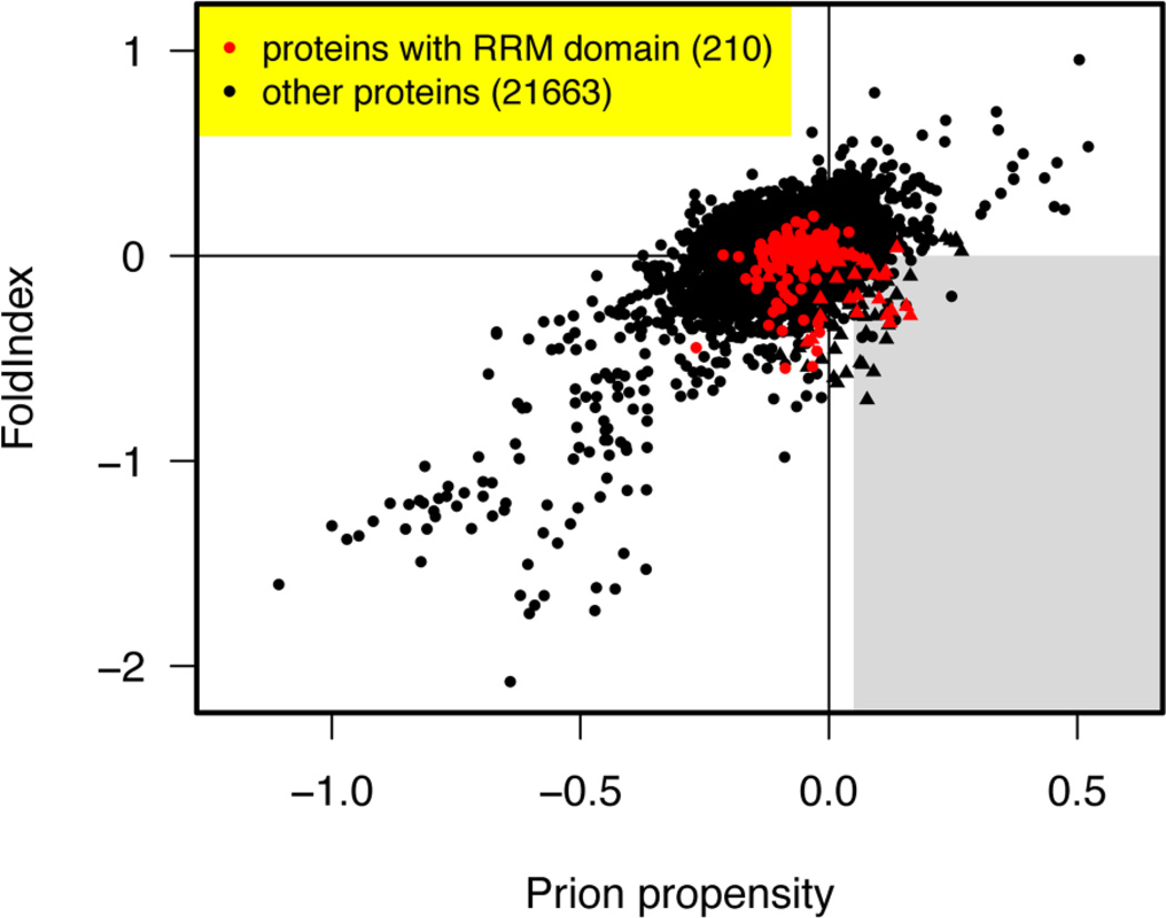
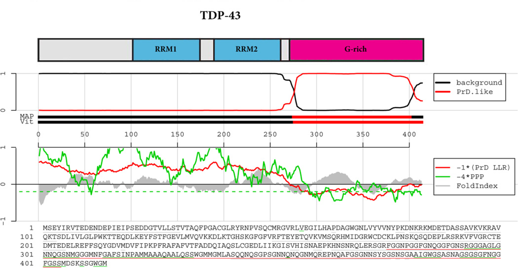
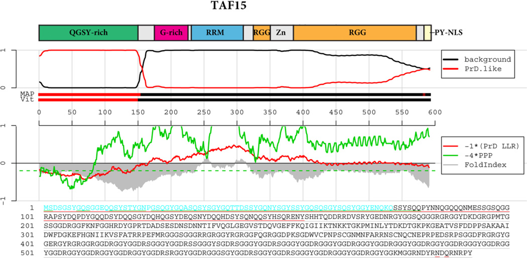
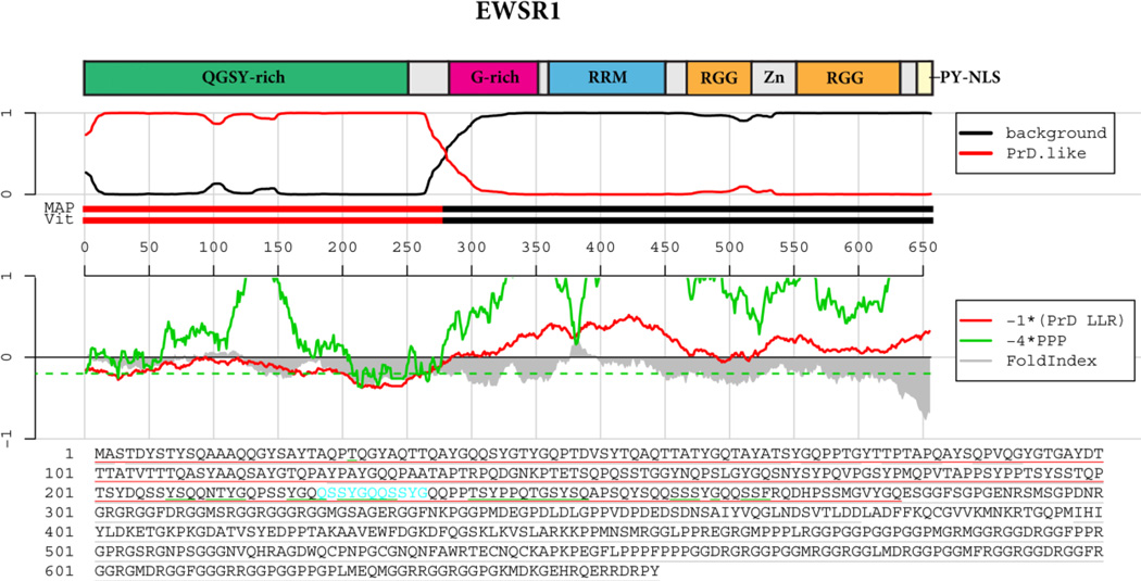
Prions are self-templating protein conformers that are naturally transmitted between individuals and promote phenotypic change. In yeast, prion-encoded phenotypes can be beneficial, neutral or deleterious depending upon genetic background and environmental conditions. A distinctive and portable 'prion domain' enriched in asparagine, glutamine, tyrosine and glycine residues unifies the majority of yeast prion proteins. Deletion of this domain precludes prionogenesis and appending this domain to reporter proteins can confer prionogenicity. An algorithm designed to detect prion domains has successfully identified 19 domains that can confer prion behavior. Scouring the human genome with this algorithm enriches a select group of RNA-binding proteins harboring a canonical RNA recognition motif (RRM) and a putative prion domain. Indeed, of 210 human RRM-bearing proteins, 29 have a putative prion domain, and 12 of these are in the top 60 prion candidates in the entire genome. Startlingly, these RNA-binding prion candidates are inexorably emerging, one by one, in the pathology and genetics of devastating neurodegenerative disorders, including: amyotrophic lateral sclerosis (ALS), frontotemporal lobar degeneration with ubiquitin-positive inclusions (FTLD-U), Alzheimer's disease and Huntington's disease. For example, FUS and TDP-43, which rank 1st and 10th among RRM-bearing prion candidates, form cytoplasmic inclusions in the degenerating motor neurons of ALS patients and mutations in TDP-43 and FUS cause familial ALS. Recently, perturbed RNA-binding proteostasis of TAF15, which is the 2nd ranked RRM-bearing prion candidate, has been connected with ALS and FTLD-U. We strongly suspect that we have now merely reached the tip of the iceberg. We predict that additional RNA-binding prion candidates identified by our algorithm will soon surface as genetic modifiers or causes of diverse neurodegenerative conditions. Indeed, simple prion-like transfer mechanisms involving the prion domains of RNA-binding proteins could underlie the classical non-cell-autonomous emanation of neurodegenerative pathology from originating epicenters to neighboring portions of the nervous system. This article is part of a Special Issue entitled RNA-Binding Proteins.


Figures

Figure 1
Figure 1. Human RNA-binding proteins with prion-like domains
All human proteins from Ensembl release GRCh37.59 (78928 proteins including variant isoforms) were scanned for prion-like domains. The FoldIndex (Prilusky et al., 2005) and prion propensity scores (Toombs et al., 2010) are plotted for each human protein. Only the highest scoring protein isoform mapping to any single Ensembl gene ID is shown. RRM-containing proteins are indicated in red, and other proteins in black. Prion candidates contain regions that satisfy both conditions in a way that places them in the grey shaded sweet spot in the lower right. Both the FoldIndex and prion propensity scores represent averages of scores for 41 consecutive 41 amino acid (AA) windows (Toombs et al., 2010). The plotted scores for each protein are based on the consecutive windows that maximize the signed distance to the boundary of the grey region, which is positive for regions satisfying both conditions and negative otherwise. Proteins containing a region with prion-like amino acid composition are indicated by triangles (Alberti et al., 2009). These are defined as positive log-likelihood ratio when averaged over the 41 consecutive windows, based on the hidden Markov model of Alberti et al. (2009) but without imposing a hard minimum length requirement of 60 residues in the Viterbi parse. The prion-like amino acid frequencies were set to the average for 19 experimentally verified prion-like domains in S. cerevisiae (Alberti et al., 2009), and the background amino acid frequencies were set to the average of the proteome-wide amino acid frequencies in S. cerevisiae and H. sapiens. The RRM proteins that satisfy the Alberti et al. (2009) criteria are listed and ranked in Table 1.
Figure 2
Figure 2. TDP-43 prion domain prediction
The top panel shows the domain architecture of TDP-43. RRM=RNA-recognition motif; G-rich=Glycine-rich domain. Below the cartoon the probability of each residue belonging to the hidden Markov model state prion domain or ‘background’ is plotted; the tracks ‘MAP’ and ‘Vit’ illustrate the Maximum a Posteriori and the Viterbi parses of the protein into the prion domain or non-prion domain (Alberti et al., 2009). The plots in the middle panel show the log-likelihood ratio scores (PrD LLR) from the Alberti et al. algorithm in red (Alberti et al., 2009), the predicted prion propensity (PPP) log-odds ratio scores from the Toombs et al. algorithm in green (Toombs et al., 2010) and FoldIndex scores in grey (Prilusky et al., 2005), each averaged over sliding windows of 41 residues. Note that the curves are rescaled to give similar ranges, and so that negative scores are suggestive of both disorder and prion propensity; the rescaled cutoff corresponding to PPP > 0.05 is indicated by the dashed green line. The lower part of the panel shows the primary sequence of TDP-43. The Alberti prion domain is underlined in red (Alberti et al., 2009), the Toombs prion domain in underlined in green (Toombs et al., 2010), and the cyan residues represent the regions that satisfy these requirements of disorder and prion propensity of the Toombs algorithm (Toombs et al., 2010) as well as the amino acid composition requirement of the Alberti algorithm (Alberti et al., 2009). Note the lack of cyan residues for TDP-43.
Figure 3
Figure 3. FUS prion-like domain prediction
The top panel shows the domain architecture of FUS. QGSY-rich=Glutamine, glycine, serine and tyrosine-rich domain; RRM=RNA-recognition motif; G-rich=Glycine-rich domain; RRM=RNA-recognition motif; RGG=RGG domain, a domain with repeated Gly-Gly dipeptides interspersed with Arg and aromatic residues. Zn=Zinc finger motif. Below the cartoon the probability of each residue belonging to the Hidden Markov Model state prion domain or ‘background’ is plotted; the tracks ‘MAP’ and ‘Vit’ illustrate the Maximum a Posteriori and the Viterbi parses of the protein into the prion domain or non-prion domain (Alberti et al., 2009). The plots in the middle panel show the log-likelihood ratio scores (PrD LLR) from the Alberti et al. algorithm in red (Alberti et al., 2009), the predicted prion propensity (PPP) log-odds ratio scores from the Toombs et al. algorithm in green (Toombs et al., 2010) and FoldIndex scores in grey (Prilusky et al., 2005), each averaged over sliding windows of residues. Note that the curves are rescaled to give similar ranges, and so that negative scores are suggestive of both disorder and prion propensity; the rescaled cutoff corresponding to PPP > 0.05 is indicated by the dashed green line. The lower part of the panel shows the primary sequence of TDP-43. The Alberti prion domain is underlined in red (Alberti et al., 2009), the centers of windows satisfying the disorder and prion propensity criteria of Toombs are underlined in grey and green (Toombs et al., 2010), and the cyan residues represent the centers of regions that satisfy both Toombs criteria as well as the amino acid composition requirement of the Alberti algorithm.
Figure 4
Figure 4. TAF15 prion-like domain prediction
The top panel shows the domain architecture of TAF15. QGSY-rich=Glutamine, glycine, serine and tyrosine-rich domain; RRM=RNA-recognition motif; G-rich=Glycine-rich domain; RRM=RNA-recognition motif; RGG=RGG domain, a domain with repeated Gly-Gly dipeptides interspersed with Arg and aromatic residues. Zn=Zinc finger motif. Below the cartoon the probability of each residue belonging to the Hidden Markov Model state prion domain or ‘background’ is plotted; the tracks ‘MAP’ and ‘Vit’ illustrate the Maximum a Posteriori and the Viterbi parses of the protein into the prion domain or non-prion domain (Alberti et al., 2009). The plots in the middle panel show the log-likelihood ratio scores (PrD LLR) from the Alberti et al. algorithm in red (Alberti et al., 2009), the predicted prion propensity (PPP) log-odds ratio scores from the Toombs et al. algorithm in green (Toombs et al., 2010) and FoldIndex scores in grey (Prilusky et al., 2005), each averaged over sliding windows of 41 residues. Note that the curves are rescaled to give similar ranges, and so that negative scores are suggestive of both disorder and prion propensity; the rescaled cutoff corresponding to PPP > 0.05 is indicated by the dashed green line. The lower part of the panel shows the primary sequence of TDP-43. The Alberti prion domain is underlined in red (Alberti et al., 2009), the centers of windows satisfying the disorder and prion propensity criteria of Toombs are underlined in grey and green (Toombs et al., 2010), and the cyan residues represent the centers of regions that satisfy both Toombs criteria as well as the amino acid composition requirement of the Alberti algorithm.
Figure 5
Figure 5. EWSR1 prion-like domain prediction
The top panel shows the domain architecture of EWSR1. QGSY-rich=Glutamine, glycine, serine and tyrosine-rich domain; RRM=RNA-recognition motif; G-rich=Glycine-rich domain; RRM=RNA-recognition motif; RGG=RGG domain, a domain with repeated Gly-Gly dipeptides interspersed with Arg and aromatic residues. Zn=Zinc finger motif. Below the cartoon the probability of each residue belonging to the Hidden Markov Model state prion domain or ‘background’ is plotted; the tracks ‘MAP’ and ‘Vit’ illustrate the Maximum a Posteriori and the Viterbi parses of the protein into the prion domain or non-prion domain (Alberti et al., 2009). The plots in the middle panel show the log-likelihood ratio scores (PrD LLR) from the Alberti et al. algorithm in red (Alberti et al., 2009), the predicted prion propensity (PPP) log-odds ratio scores from the Toombs et al. algorithm in green (Toombs et al., 2010) and FoldIndex scores in grey (Prilusky et al., 2005), each averaged over sliding windows of 41 residues. Note that the curves are rescaled to give similar ranges, and so that negative scores are suggestive of both disorder and prion propensity; the rescaled cutoff corresponding to PPP > 0.05 is indicated by the dashed green line. The lower part of the panel shows the primary sequence of TDP-43. The Alberti prion domain is underlined in red (Alberti et al., 2009), the centers of windows satisfying the disorder and prion propensity criteria of Toombs are underlined in grey and green (Toombs et al., 2010), and the cyan residues represent the centers of regions that satisfy both Toombs criteria as well as the amino acid composition requirement of the Alberti algorithm.

References

    1. Aguzzi A. Cell biology: Beyond the prion principle. Nature. 2009;459:924–925. - PubMed
    1. Aguzzi A, Rajendran L. The transcellular spread of cytosolic amyloids, prions, and prionoids. Neuron. 2009;64:783–790. - PubMed
    1. Al-Sarraj S, King A, Troakes C, Smith B, Maekawa S, Bodi I, Rogelj B, Al-Chalabi A, Hortobagyi T, Shaw CE. p62 positive, TDP-43 negative, neuronal cytoplasmic and intranuclear inclusions in the cerebellum and hippocampus define the pathology of C9orf72-linked FTLD and MND/ALS. Acta Neuropathol. 2011;122:691–702. - PubMed
    1. Alberti S, Halfmann R, King O, Kapila A, Lindquist S. A systematic survey identifies prions and illuminates sequence features of prionogenic proteins. Cell. 2009;137:146–158. - PMC - PubMed
    1. Arai T, Hasegawa M, Akiyama H, Ikeda K, Nonaka T, Mori H, Mann D, Tsuchiya K, Yoshida M, Hashizume Y, Oda T. TDP-43 is a component of ubiquitin-positive tau-negative inclusions in frontotemporal lobar degeneration and amyotrophic lateral sclerosis. Biochem Biophys Res Commun. 2006;351:602–611. - PubMed

Publication types

MeSH terms