

Mapping membrane protein structure with fluorescence
- PMID: 22445227
- PMCID: PMC3498957
- DOI: 10.1016/j.sbi.2012.02.004
Mapping membrane protein structure with fluorescence
Abstract
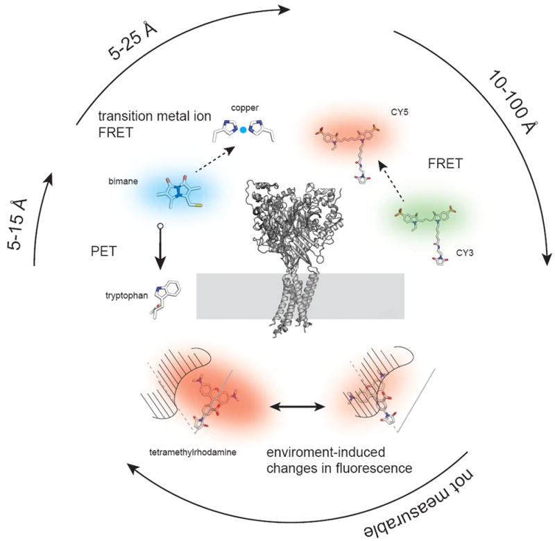
Membrane proteins regulate many cellular processes including signaling cascades, ion transport, membrane fusion, and cell-to-cell communications. Understanding the architecture and conformational fluctuations of these proteins is critical to understanding their regulation and functions. Fluorescence methods including intensity mapping, fluorescence resonance energy transfer (FRET), and photo-induced electron transfer, allow for targeted measurements of domains within membrane proteins. These methods can reveal how a protein is structured and how it transitions between different conformational states. Here, I will review recent work done using fluorescence to map the structures of membrane proteins, focusing on how each of these methods can be applied to understanding the dynamic nature of individual membrane proteins and protein complexes.
Published by Elsevier Ltd.
Figures
References
-
- Long SB, Tao X, Campbell EB, MacKinnon R. Atomic structure of a voltage-dependent K+ channel in a lipid membrane-like environment. Nature. 2007;450:376–382. - PubMed
-
- Hille B. Ion channels of excitable membranes. 3. Sunderland, Mass: Sinauer; 2001.
Publication types
MeSH terms
Substances
Grants and funding
LinkOut - more resources
Full Text Sources
