Review
. 2012 Mar 22;73(6):1068-81.
doi: 10.1016/j.neuron.2012.03.005. Epub 2012 Mar 21.

Growth cone travel in space and time: the cellular ensemble of cytoskeleton, adhesion, and membrane

Affiliations
Review

Growth cone travel in space and time: the cellular ensemble of cytoskeleton, adhesion, and membrane

Eric A Vitriol et al. Neuron. .

Abstract

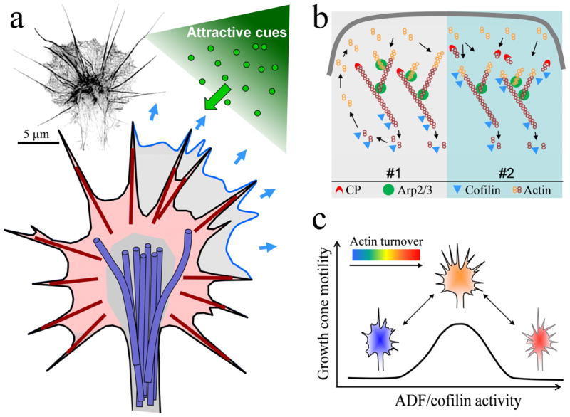
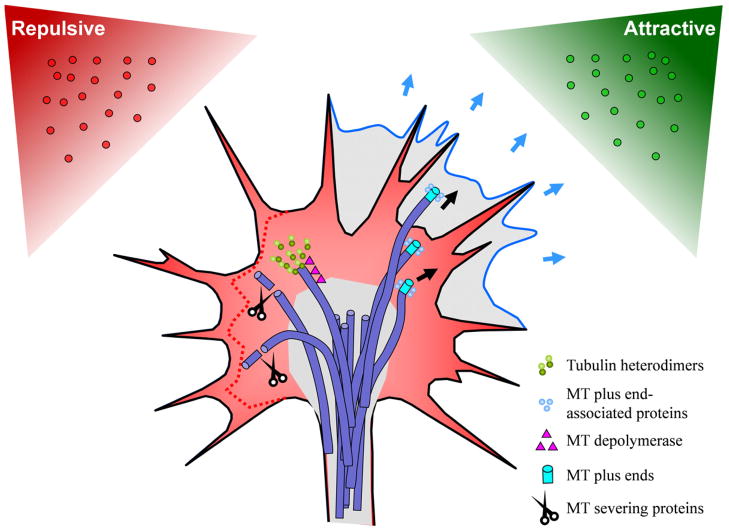
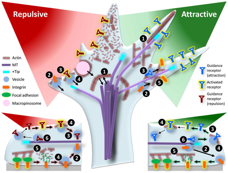
Growth cones, found at the tip of axonal projections, are the sensory and motile organelles of developing neurons that enable axon pathfinding and target recognition for precise wiring of the neural circuitry. To date, many families of conserved guidance molecules and their corresponding receptors have been identified that work in space and time to ensure billions of axons to reach their targets. Research in the past two decades has also gained significant insight into the ways in which growth cones translate extracellular signals into directional migration. This review aims to examine new progress toward understanding the cellular mechanisms underlying directional motility of the growth cone and to discuss questions that remain to be addressed. Specifically, we will focus on the cellular ensemble of cytoskeleton, adhesion, and membrane and examine how the intricate interplay between these processes orchestrates the directed movement of growth cones.


Figures

Figure 1
Figure 1
Actin cytoskeleton of the growth cone. (a) Schematic of the actin cytoskeleton of a growth cone undergoing an attractive guidance response. The growth cone’s periphery contains actin-rich lamellipodia (light red shaded) and filopodia (dark red lines. The lamellipodia consists of a network of short, branched actin filaments that serves as the protrusion machinery of the growth cone. Newly formed lamellipodia on the side undergoing a positive turning response is shown in blue. Filopodia are composed of long, bundles actin filaments. They participate in environment sensing and guidance. Microtubules in the growth cone (shown in purple) are largely restricted to the central region (gray shaded) by the actin cytoskeleton. The inset on the top left shows a fluorescent image of the actual F-actin architecture within the growth cone. This image was obtained using the Nikon N-SIM Super Resolution microscope and inverted in grayscale for display. Scale bar: 5 μm. (b) Two models of ADF/cofilin-mediated regulation of actin dynamics underlying lamellipodial protrusion. The #1 is the classical model in which ADF/cofilin functions mainly in depolymerization at the rear of the actin meshwork and recycling the actin monomers to the leading front for assembly. In the #2 model, ADF/cofilin severing creates new barbed ends to promote actin assembly and membrane protrusion. (c) A hypothesized model in which an optimal range of ADF/cofilin activity may be required for growth cone motility. Above and below this range of ADF/cofilin activity inhibits growth cone motility. Depending on upon the amount of active ADF/cofilin and the dynamic state of the actin network, a gradient of guidance cues could asymmetrically target the ADF/cofilin activity to generate either attractive (into the optimal range) or repulsive (out of the optimal range) responses.
Figure 2
Figure 2
Microtubules in growth cone steering. This schematic shows the hypothesized model involving asymmetric modification of MT dynamics during growth cone attraction and repulsion. Newly formed lamellipodia is shown in blue, retracting lamellipodia is indicated by the dotted red line. Microtubules in the growth cone (shown in purple) are largely restricted to the Central region by the actin cytoskeleton, but some enter into the P-region, where they play an important role in axon guidance. MT localization is controlled by the actin cytoskeleton and a host of MT regulatory proteins. During retraction, MTs are removed from the periphery through selective targeting by depolymerases and severing proteins. During attraction, proteins bind to MT plus-ends, stabilizing them or enhancing polymerization and further growth.
Figure 3
Figure 3
Schematic showing the complexity of cross-talk among different cellular machineries. The directional movement of the growth cone involves membrane protrusion at the front and retraction at the rear, which are controlled by bi-directional interactions between the actin and microtubules (1). Successful locomotion also requires the formation of new adhesion at the front and the destruction of adhesion at the rear, which are mediated by membrane recycling (2). Vesicular trafficking and recycling also control and regulate the number and distribution of guidance receptors on the surface, which will impact the spatiotemporal signal transduction (3). While the actin cytoskeleton is the driving force for membrane protrusion, it also regulates endocytosis and exocytosis in a spatiotemporal fashion, which could impact both the adhesion and receptor recycling (4). The actin cytoskeleton is also a part of the integrin-adhesion complex and regulates the stability and turnover of adhesion (5). Finally, microtubules and their plus-ends play a role in trafficking membrane channels and receptors (6).

References

    1. Aizawa H, Wakatsuki S, Ishii A, Moriyama K, Sasaki Y, Ohashi K, Sekine-Aizawa Y, Sehara-Fujisawa A, Mizuno K, Goshima Y, Yahara I. Phosphorylation of cofilin by LIM-kinase is necessary for semaphorin 3A-induced growth cone collapse. Nat Neurosci. 2001;4:367–373. - PubMed
    1. Akin O, Mullins RD. Capping protein increases the rate of actin-based motility by promoting filament nucleation by the Arp2/3 complex. Cell. 2008;133:841–851. - PMC - PubMed
    1. Andrianantoandro E, Pollard TD. Mechanism of actin filament turnover by severing and nucleation at different concentrations of ADF/cofilin. Mol Cell. 2006;24:13–23. - PubMed
    1. Arakawa Y, Bito H, Furuyashiki T, Tsuji T, Takemoto-Kimura S, Kimura K, Nozaki K, Hashimoto N, Narumiya S. Control of axon elongation via an SDF-1alpha/Rho/mDia pathway in cultured cerebellar granule neurons. J Cell Biol. 2003;161:381–391. - PMC - PubMed
    1. Aratyn-Schaus Y, Gardel ML. Transient frictional slip between integrin and the ECM in focal adhesions under myosin II tension. Curr Biol. 2010;20:1145–1153. - PMC - PubMed

Publication types

LinkOut - more resources