. 2012 Mar 22;73(6):1116-26.
doi: 10.1016/j.neuron.2012.02.009. Epub 2012 Mar 21.

Sonic hedgehog expression in corticofugal projection neurons directs cortical microcircuit formation

Affiliations

Sonic hedgehog expression in corticofugal projection neurons directs cortical microcircuit formation

Corey C Harwell et al. Neuron. .

Abstract

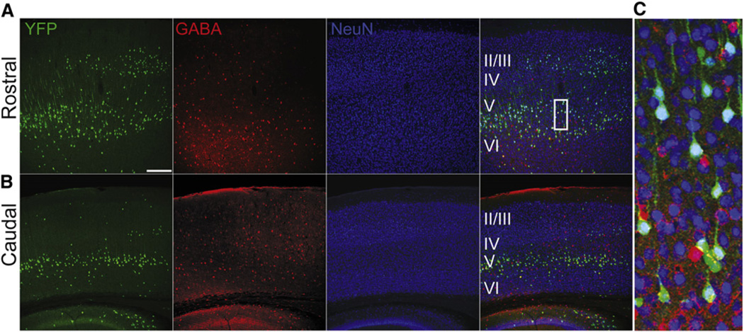
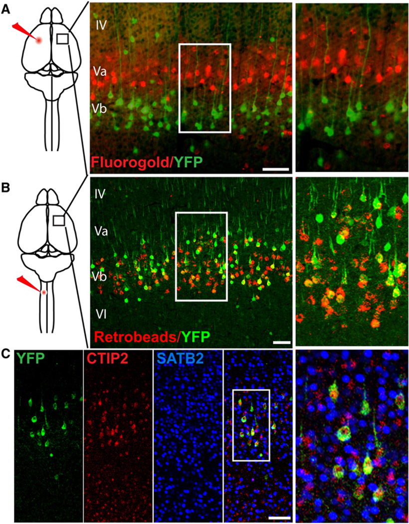
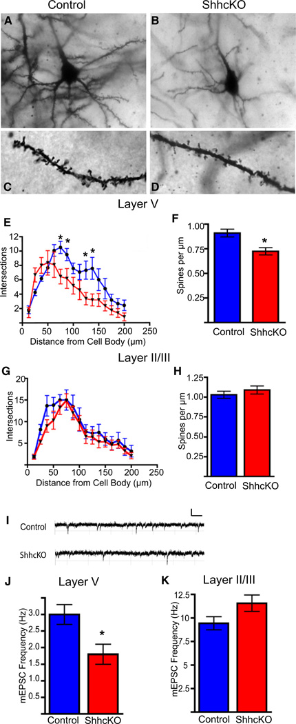
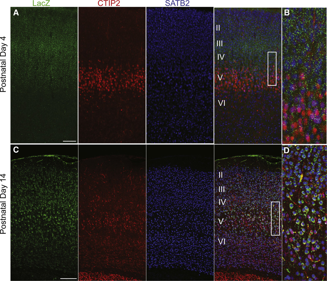
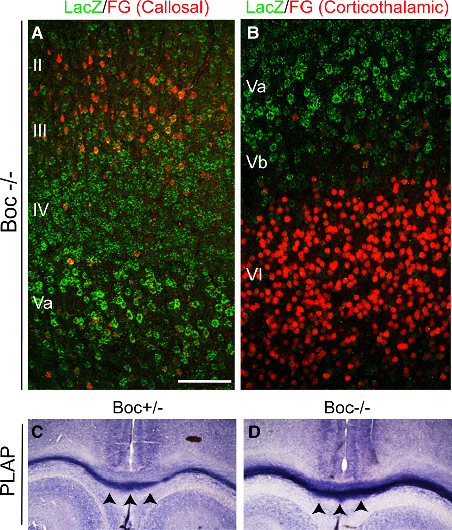
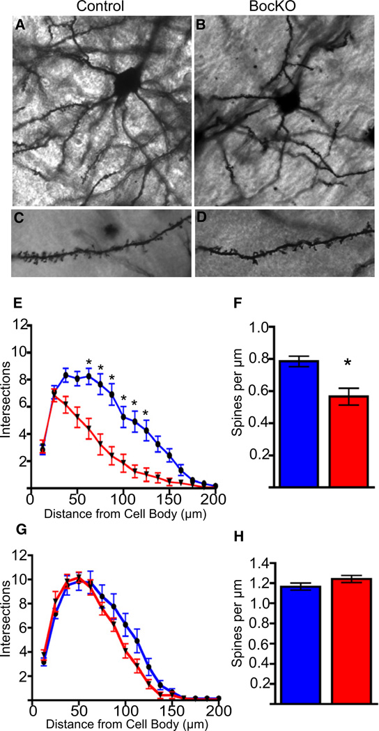
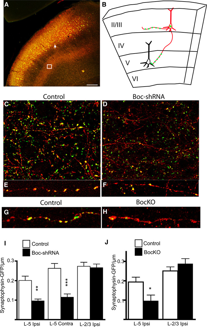
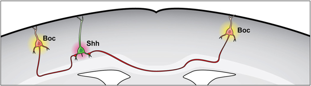
The precise connectivity of inputs and outputs is critical for cerebral cortex function; however, the cellular mechanisms that establish these connections are poorly understood. Here, we show that the secreted molecule Sonic Hedgehog (Shh) is involved in synapse formation of a specific cortical circuit. Shh is expressed in layer V corticofugal projection neurons and the Shh receptor, Brother of CDO (Boc), is expressed in local and callosal projection neurons of layer II/III that synapse onto the subcortical projection neurons. Layer V neurons of mice lacking functional Shh exhibit decreased synapses. Conversely, the loss of functional Boc leads to a reduction in the strength of synaptic connections onto layer Vb, but not layer II/III, pyramidal neurons. These results demonstrate that Shh is expressed in postsynaptic target cells while Boc is expressed in a complementary population of presynaptic input neurons, and they function to guide the formation of cortical microcircuitry.


Figures

Figure 1
Figure 1. Sonic Hedgehog Is Expressed Primarily in Deep-Layer Cortical Pyramidal Neurons
(A–C) Immunohistochemistry of coronal sections of adult (8 weeks) ShhCre; RYFP brains, located in both the (A) rostral and (B) caudal axis of the brain. Ninety-six percent YFP+ cells (green) are NeuN positive (blue), while only 4% of cells are also positive for GABA (red), indicating that the vast majority of Shh expressing cells in the cortex are cortical pyramidal neurons. n = 3 animals, 812 cells. Scale bars represent 250 µm (A and B).
Figure 2
Figure 2. Sonic Hedgehog Is Expressed Exclusively in Corticofugal Projection Neurons
(A) Schematic of fluorogold (red) injection into the cortex of ShhGFPCre; RYFP mice. A coronal section showing no overlap between fluorogold labeled callosal projections (red) and YFP+ (green) Shh lineage neurons. (B) In contrast, when fluorescent retrobeads (red) were injected into the corticospinal tract, 60% (n = 2 animals, 612 cells) of bead positive cells were YFP+ (green). (C) P14 ShhGFPCre; RYFP mice were immunostained for the corticofugal projection marker CTIP2 (red) and 92% (n = 2 animals, 200 cells) YFP+ cells were CTIP2+, while none of the cells were labeled with the callosal projection marker SATB2 (blue). Scale bars represent 150 µm.
Figure 3
Figure 3. Sonic Hedgehog Is Required for Proper Synaptic Development of Deep-Layer Cortical Neurons In Vivo
(A–D) Golgi stained control and ShhcKO (Emx1;Shhfl/fl) brains show decreased branching and dendritic complexity and spine density of layer V neurons in Shh conditional knockouts compared to controls. (E) Sholl analysis of layer V neurons reveals significantly fewer intersections of dendritic processes at 75, 87.5, 125, and 137.5 µm from the cell soma. (F) There is also significant reduction in basal dendritic spine density in the ShhcKO animals when compared to the controls (0.91 ± 0.04 versus 0.72 ± 0.04). (G and H) There is no significant change in dendritic branching or spine density in neurons of the superficial cortical layers. (I–K) Recordings of mEPSCs from layer V neurons reveals a significant decrease in mEPSC frequency in Shh conditional knockouts (1.8 ± 0.3 Hz) when compared to littermate controls (3.0 ± 0.3 Hz). There is no significant change in mini frequency observed in superficial layer neurons (5.34 Hz versus 5.36 Hz), n = 3 animals per group and 9 cells per group. *p < 0.01, t test.
Figure 4
Figure 4. The Sonic Hedgehog Receptor Boc Is Expressed Primarily by SATB2-Positive Callosal Projection Neuron Subtypes
(A) Postnatal day 3 coronal sections from Boc+/− LacZ-ires-PLAP mice showing LacZ expression primarily in SATB2 positive neurons located in layers II, III, IV, and Va. (B) Higher, magnification view of the box area in (A). (C) Postnatal day 14 coronal sections from Boc+/− LacZ-ires-PLAP mice showing LacZ expression primarily in SATB2 positive neurons located in layers II, III, IV, and Va. (D) Higher magnification of the boxed area in (C). Note the increased intensity of LacZ staining in at P14. Scale bars represent 100 µm (A); 250 µm (B).
Figure 5
Figure 5. Boc Is Not Required for Long-Range Callosal Neuron Axon Guidance
(A) Fluorogold (FG) retrograde labeling of callosal projection neurons located in layers II/III in homozygous Boc mutant mice showing that axons still reach the contralateral cortex. (B) Fluorogold retrograde labeling of corticothalamic projection neurons located primarily in cortical layer VI in homozygous Boc mutant mice, showing that the boundary of expression and maintenance of projection identity is maintained in the mutants. (C and D) PLAP staining of callosal axon projections in (C) heterozygous and (D) homozygous Boc mutants. Black arrowheads indicate the corpus callosum. Note the darker PLAP staining in the homozygote due to the additional copy of the PLAP gene.
Figure 6
Figure 6. Loss of Boc Function Reduces Dendritic Growth and Spine Density of Deep-Layer Cortical Neurons In Vivo
(A–D) P21 Golgi stained control and BocKO brains show that there is decreased branching, dendritic complexity, and spine density of layer V neurons when compared to control animals. (E) Sholl analysis of layer V neurons reveals significantly fewer intersections of dendritic processes between 72 to 120 µm from the cell soma. (F) There is also a significant reduction in the density of basal dendritic spines of layer V neurons (0.78 ± 0.03 versus 0.57 ± 0.05). (G and H) Sholl analysis and spine counts in layer II/III did not reveal significant differences between groups.
Figure 7
Figure 7. Layer II/III Boc Knockdown Leads to Loss of Synaptophysin Puncta in Layer V
(A) Coronal section of a P28 brain coelectroporated at E14 with a control or BocshRNA vector, pCAG-H2B-GFP-2A-myr-Tdtomato and a synaptophysin-GFP plasmid. The white arrow indicates H2B-GFP nuclei of electroporated cells while membrane Tdtomato/Synaptophysin-GFP labeled axons concentrated in layer V are highlighted inside the white box. (B) Schematic of an electroporated cell with red axons in layers II/III and V, and dots of synaptophysin-GFP puncta along the axon. (C–F) Images of axons (red) and synaptophysin-GFP puncta (green) in layer V of control and Boc-shRNA electroporated cells. (G and H) Images of layer V axons from P14 control and BocKO animals. (I) There is a significant reduction in the density of GFP puncta from axons located in both ipsilateral and contralateral layer V (0.1 ± 0.01 and 0.11 ± 0.02, respectively, n = 80 axon segments) when compared with control hairpin electroporated (0.2 ± 0.02 and 0.26 ± 0.03 ±, n = 63 axon segments) cells. There is no significant difference in density of axons located in layer II/III (0.27 ± 0.02 and 0.26 ± 0.02, n = 37 and n = 29 axon segments). (J) There is a similar pattern of reduced synaptophysin-GFP puncta in layer V of BocKO animals when compared with controls (0.10 ± 0.03 and 0.19 ± 0.02, n = 15 axon segments for each group), while layer II/III axons were not significantly different (0.29 ± 0.03 and 0.25 ± 0.02, n = 15 axon segments for each group). *p < 0.05, **p < 0.01, ***p < 0.001, t test. Scale bars represent 200 µm.
Figure 8
Figure 8. Boc Is Required for Proper Layer III to V Cortical Circuit Formation
(A) Schematic of coelectroporation of hairpin plasmids with a channelrhodopsin-2 (ChR2) vector where (red) electroporated cells in layer II/III are activated with blue light while responses of (black) unelectroporated cells in layer II/III are recorded. (B) Voltage clamp recordings of the responses of unelectroporated cells located in layers II/III, and a recording from ChR2 electroporated cells showing normal spiking when flashed with blue light. (C) Schematic of coelectroporation of hairpin plasmids with a channelrhodopsin-2 (ChR2) vector where (red) electroporated cells in layer II/III are activated with blue light while responses of (black) unelectroporated cells in layer V are recorded. (D) Voltage clamp recordings of the responses of unelectroporated cells located in layer V. (E) There is no significant change in the response amplitude of layer II/III neurons of control (587.7 ± 90.1 pA) and Boc-shRNA (852.2 ± 180.4 pA) or BocHet (1,165.7 ± 412.8 pA) and BocKO (2,002.2 ± 585.3 pA). n = 4 animals, and 4–10 cells per group (see Experimental Procedures for more details). **p < 0.01, *p < 0.05 one-way ANOVA. (CTL HP, ShhcKO, BocHP) unpaired t test (BocKO and BocHet) Scale bars represent 200 pA and 40 ms. (F) EPSC amplitude is significantly reduced in layer V cells of Boc-shRNA, BocKO, and ShhcKO (51.3 ± 14.3 and 180.4 ± 73.0 pA and 58.3 ± 6.5.0 pA, respectively) when compared to the control condition (1,176.0 ± 386.0 pA) or Boc heterozygous animals (820 ± 291.0 pA). Scale bars represent 100 pA and 40 ms.
Figure 9
Figure 9. Complementary Expression of Sonic Hedgehog and Boc Directs Synapse Formation
Sonic Hedgehog expression by corticofugal neurons directs cortical circuit formation with local/callosal Boc expressing cells. Model illustrates the cell type-specific expression of the ligand Sonic Hedgehog and its receptor Boc into distinct nonoverlapping populations of projection neurons. Sonic Hedgehog is expressed by corticofugal neurons, while Boc is expressed by local and callosal projection neurons. Sonic Hedgehog released by corticofugal neurons functions to guide the formation of cortical microcircuitry with Boc expressing neurons.

Comment in

References

    1. Ahn S, Joyner AL. In vivo analysis of quiescent adult neural stem cells responding to Sonic hedgehog. Nature. 2005;437:894–897. - PubMed
    1. Alcamo EA, Chirivella L, Dautzenberg M, Dobreva G, Fariñas I, Grosschedl R, McConnell SK. Satb2 regulates callosal projection neuron identity in the developing cerebral cortex. Neuron. 2008;57:364–377. - PubMed
    1. Anderson CT, Sheets PL, Kiritani T, Shepherd GMG. Sublayer-specific microcircuits of corticospinal and corticostriatal neurons in motor cortex. Nat. Neurosci. 2010;13:739–744. - PMC - PubMed
    1. Arlotta P, Molyneaux BJ, Chen J, Inoue J, Kominami R, Macklis JD. Neuronal subtype-specific genes that control corticospinal motor neuron development in vivo. Neuron. 2005;45:207–221. - PubMed
    1. Britanova O, de Juan Romero C, Cheung A, Kwan KY, Schwark M, Gyorgy A, Vogel T, Akopov S, Mitkovski M, Agoston D, et al. Satb2 is a postmitotic determinant for upper-layer neuron specification in the neocortex. Neuron. 2008;57:378–392. - PubMed

Publication types

MeSH terms

Substances