. 2012 Apr 4;15(4):545-53.
doi: 10.1016/j.cmet.2012.01.022. Epub 2012 Mar 22.

Macrophage autophagy plays a protective role in advanced atherosclerosis

Affiliations

Macrophage autophagy plays a protective role in advanced atherosclerosis

Xianghai Liao et al. Cell Metab. .

Abstract

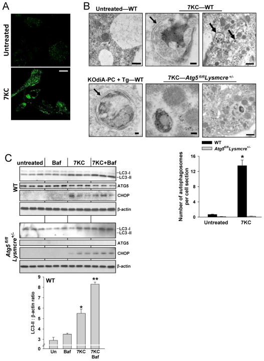
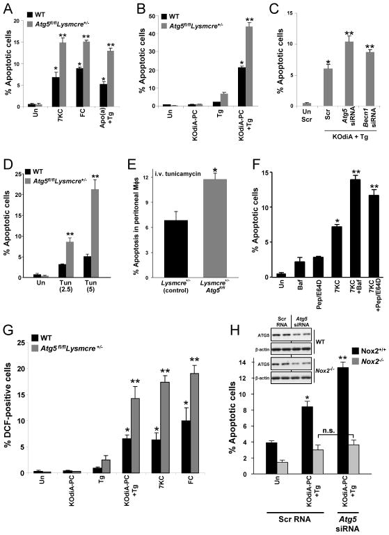
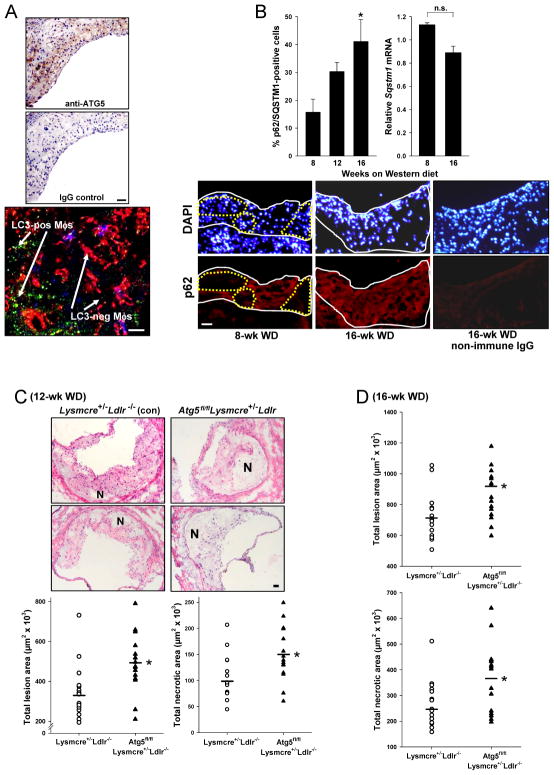
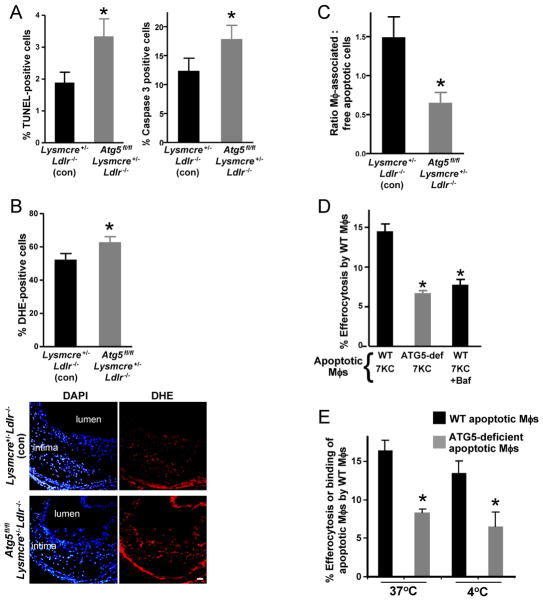
In advanced atherosclerosis, macrophage apoptosis coupled with defective phagocytic clearance of the apoptotic cells (efferocytosis) promotes plaque necrosis, which precipitates acute atherothrombotic cardiovascular events. Oxidative and endoplasmic reticulum (ER) stress in macrophages are important causes of advanced lesional macrophage apoptosis. We now show that proapoptotic oxidative/ER stress inducers trigger another stress reaction in macrophages, autophagy. Inhibition of autophagy by silencing ATG5 or other autophagy mediators enhances apoptosis and NADPH oxidase-mediated oxidative stress while at the same time rendering the apoptotic cells less well recognized by efferocytes. Most importantly, macrophage ATG5 deficiency in fat-fed Ldlr(-/-) mice increases apoptosis and oxidative stress in advanced lesional macrophages, promotes plaque necrosis, and worsens lesional efferocytosis. These findings reveal a protective process in oxidatively stressed macrophages relevant to plaque necrosis, suggesting a mechanism-based strategy to therapeutically suppress atherosclerosis progression and its clinical sequelae.


Figures

Figure 1
Figure 1. Autophagy is Induced in Macrophages Exposed to Atherosclerosis-Related Stimulators of Apoptosis
(A) Confocal fluorescence microscopy of macrophages from GFP-LC3 transgenic mice that were left untreated or treated for 8 h with 35 μM 7-ketocholesterol (7KC). Bar = 10 μm. (B) Electron microscopy of macrophages from WT or Atg5fl/flLysmCre+/− mice that were left untreated or treated for 6 h with 7KC or 50 μg/ml of KOdiA-PC plus 0.5 μM thapsigargin (KOdiA-PC + Tg). Double-membranes are depicted by arrows. Bar, 100 nm for left and middle row of images and 500 nm for right row of images. Quantification of the average number of autophagosomes per cell section in untreated and 7KC-treated WT and Atg5fl/flLysmCre+/− macrophages is shown (mean ± S.E.M.; *p < 0.01 vs. other groups; n = 10 cell sections/group). (C) Macrophages from WT or Atg5fl/flLysmCre+/− mice were left untreated or incubated for 4 h with 7KC. In some groups, 10 μM bafilomycin A1 (Baf) was added during the last 2 h. Cell extracts were probed for LC3-I, LC3-II, Atg5, CHOP, and β-actin by immunoblot. Densitometric quantification of the WT immunoblot data are shown in the graph (mean ± S.E.M.; *,**p < 0.05 vs. the other groups and each other).
Figure 2
Figure 2. Inhibition of Autophagy Increases Apoptosis and NADPH Oxidase-Mediated Oxidative Stress
For (A) – (D), macrophages from WT or Atg5fl/flLysmCre+/− mice were left untreated (Un) or incubated as below and then assayed for apoptosis by annexin V staining (mean ± S.E.M.; *p < 0.05, **p < 0.01 vs. the other groups). (A) Incubation times were 20 h for 7KC, 18 h for free cholesterol (FC)-loading, and 20 h for 50 μg/ml apolipoprotein(a) [Apo(a)] plus 0.5 μM thapsigargin (Tg). (B) Incubation time was 24 h for 50 μg/ml of KOdiA-PC alone, 0.5 μM thapsigargin (Tg) alone, or both reagents together. (C) As in (B), but the cells were pre-treated for 90 h with either scrambled RNA (Scr) or siRNA targeting Atg5 or Becn1. (D) Incubation times were 24 h for 2.5 or 5.0 μg/ml tunicamycin (Tun). (E) Lysmcre+/− (control) and Atg5fl/flLysmcre+/− mice were injected i.v. with 0.02 mg/kg tunicamycin, and 16 h later freshly harvested peritoneal macrophages were assayed for apoptosis using annexin V (mean ± S.E.M.; *p < 0.05). (F) Incubation time was 20 h for 7KC in the absence or presence of either 20 nM bafilomycin A1 or 10 μg/ml of pepstatin plus 10 μg/ml of E64D. (G) Macrophages from WT or Atg5fl/flLysmcre+/− mice were left untreated or incubated with the indicated reagents as above. The cells were then stained with DCFDA, viewed by fluorescence microscopy, and quantified for the percent of DCF-positive cells (mean ± S.E.M.; *p < 0.05, **p < 0.01 vs. the other groups). (H) Macrophages from Nox2+/+ or Nox2−/− mice were left untreated or incubated with KOdiA-PC plus thapsigargin and then assayed for apoptosis, except some of the cells were pre-treated for 90 h with scrambled RNA or Atg5 siRNA. The inset shows the immunoblot of ATG5 and β-actin in macrophages treated with scrambled RNA or Atg5 siRNA (mean ± S.E.M.; *p < 0.05, **p < 0.01 vs. the other groups; n.s. = not significant).
Figure 3
Figure 3. Defective Macrophage Autophagy Promotes Plaque Necrosis in Advanced Atherosclerotic Lesions of Western diet (WD)-Fed Ldlr−/− Mice
(A) Female GFP-LC3-Ldlr−/− mice were fed the WD for 12 wks. Cross-sections of an aortic root lesion were immunostained with anti-ATG5 or control IgG (top images; bar = 20 μm) or with anti-F4/80 antibody and viewed by confocal fluorescence microscopy (bottom image; red = F4/80, green = GFP-LC3; bar = 10 μm). (B) Female Lysmcre+/−Ldlr−/− (control) and Atg5fl/flLysmcre+/−Ldlr−/− mice were fed the WD for 8, 12, or 16 wks (n = 5 per group). Lesions were then immunostained for p62/SQSTM1 and DAPI (nuclei), and lesional Sqstm1 mRNA was assayed by LCM-RT-qPCR (mean ± S.E.M.; *p = 0.02 vs. 8-wk value; n.s., not significant). Representative images for the p62 immunostaining data are shown for the 8- and 16-wk lesions, along with a non-immune control image for a 16-wk lesion. Bar, 20 μm. In these images, the intima, which is composed mostly of macrophages, is outlined by the solid white lines, and areas of the 8-wk intima that have cells negative for p62 are outlined by the dotted yellow line. (C–D) Aortic root sections of female Lysmcre+/−Ldlr−/− (control) and Atg5fl/flLysmcre+/−Ldlr−/− mice fed the WD for 12 or 16 wks were stained with H&E and then quantified for lesion and necrotic area. Representative images are shown for the 12-wk groups. N, necrotic area; Bar, 20 μm (mean ± S.E.M.; *p < 0.05; n con/experimental = 15/15 for the 12-wk cohort and 17/15 for the 16-wk cohort).
Figure 4
Figure 4. Defective Macrophage Autophagy Promotes Apoptosis, Oxidative Stress, and Defective Efferocytosis
(A–B) Sections from the 12-wk cohort in Fig.3 were stained for TUNEL, activated caspase-3, or DHE and then counterstained with DAPI (nuclei) and quantified (mean ± S.E.M.; *p < 0.05; n = 15 con/15 experimental). Bar = 20 μm. (C) The lesions were stained for TUNEL and for F4/80 (macrophages), and the ratio of TUNEL-positive cells that were F4/80-associated or not (“free”) was quantified (mean ± S.E.M.; *p < 0.05; n = 15 con/15 experimental). (D) Macrophages from WT or Atg5fl/flLysmCre+/− mice were rendered apoptotic using 7KC; one set of WT cells was also treated with bafilomycin A1 (Baf). These cells were then added to WT macrophages, and efferocytosis was assayed and expressed as the percent of WT macrophages with ingested apoptotic cells (mean ± S.E.M.; *p < 0.01). (E) The experiment labeled “37°C” was similar to the 1st and 2nd experimental groups in panel D. The experiment labeled “4°C” assayed the binding of WT and Atg5fl/flLysmCre+/− apoptotic macrophages to WT macrophages (mean ± S.E.M.; *p < 0.01).

Comment in

References

    1. Ball RY, Stowers EC, Burton JH, Cary NR, Skepper JN, Mitchinson MJ. Evidence that the death of macrophage foam cells contributes to the lipid core of atheroma. Atherosclerosis. 1995;114:45–54. - PubMed
    1. Bjorkoy G, Lamark T, Pankiv S, Overvatn A, Brech A, Johansen T. Monitoring autophagic degradation of p62/SQSTM1. Methods Enzymol. 2009;452:181–197. - PubMed
    1. Clarke R, Peden JF, Hopewell JC, Kyriakou T, Goel A, Heath SC, Parish S, Barlera S, Franzosi MG, Rust S, Bennett D, Silveira A, Malarstig A, Green FR, Lathrop M, Gigante B, Leander K, de FU, Seedorf U, Hamsten A, Collins R, Watkins H, Farrall M. Genetic variants associated with Lp(a) lipoprotein level and coronary disease. N Engl J Med. 2009;361:2518–2528. - PubMed
    1. Clausen BE, Burkhardt C, Reith W, Renkawitz R, Forster I. Conditional gene targeting in macrophages and granulocytes using LysMcre mice. Transgenic Res. 1999;8:265–277. - PubMed
    1. De Meyer GR, Martinet W. Autophagy in the cardiovascular system. Biochim Biophys Acta. 2009;1793:1485–1495. - PubMed

Publication types