Review
. 2012 Aug;22(4):693-703.
doi: 10.1016/j.conb.2012.02.009. Epub 2012 Mar 23.

Dynamical changes in neurological diseases and anesthesia

Affiliations
Review

Dynamical changes in neurological diseases and anesthesia

Michelle M McCarthy et al. Curr Opin Neurobiol. 2012 Aug.

Abstract

Dynamics of neuronal networks can be altered in at least two ways: by changes in connectivity, that is, the physical architecture of the network, or changes in the amplitudes and kinetics of the intrinsic and synaptic currents within and between the elements making up a network. We argue that the latter changes are often overlooked as sources of alterations in network behavior when there are also structural (connectivity) abnormalities present; indeed, they may even give rise to the structural changes observed in these states. Here we look at two clinically relevant states (Parkinson's disease and schizophrenia) and argue that non-structural changes are important in the development of abnormal dynamics within the networks known to be relevant to each disorder. We also discuss anesthesia, since it is entirely acute, thus illustrating the potent effects of changes in synaptic and intrinsic membrane currents in the absence of structural alteration. In each of these, we focus on the role of changes in GABAergic function within microcircuits, stressing literature within the last few years.


Figures

Figure 1
Figure 1. A prominent beta oscillation emerges in the basal ganglia and cortex of Parkinson’s disease patients
(a) Schematic diagram of some of the major connections between the nuclei of the cortico-basal ganglia-thalamic loop (note: not all connections are shown). Excitatory connections are denoted by a red arrow and inhibitory connections are denoted by a blue arrow. (b, c) Highly schematic diagrams of striatal microcircuits of possible importance in the generation of rhythmic activity in Parkinson’s disease. (b) Model networks of medium spiny neurons (MSNs) connected to each other via GABAa synapses can produce beta oscillations. Beta oscillations are amplified in the presence of high ACh, which lowers the MSN M-current conductance and increases the excitability of the individual MSNs [13••]. Lowering the maximal MSN M-current conductance is sufficient to amplify beta oscillations in the model MSN network. (c) Model networks of MSNs and fast-spiking interneurons show increased synchrony as additional FS-MSN connections are added [11•]. (d) Computational modeling of networks of striatal MSNs suggests that lower M-current conductance due to high ACh (the ‘parkinsonian’ state) can induce MSNs to spike more synchronously, as seen in the raster plots (top row) producing a beta frequency rhythm seen in the model spectrogram (second row). Experimental testing of the computational model revealed that the striatum of normal mice produces a beta frequency rhythm seen in the spectrogram (third row) and the LFP trace (last row) in the presence of the cholinergic agonist, carbachol. Subfigure d adapted from McCarthy et al. [13••].
Figure 2
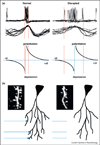
Figure 2. Altered interneuron recruitment disrupts gamma rhythm-associated spike timing and thus functional and anatomical plasticity
(a) Example of precise spike timing in principal cells afforded by normal gamma rhythms (left) and gamma rhythms disrupted by genetic manipulation of interneuron glutamatergic excitation (right). Data show local field potential triggered averages of concurrent field and intracellular records (data adapted from Fuchs et al. [86]). Below, cartoons of the standard spike timing-dependent plasticity (STDP) curves show the marked discontinuity around 0 ms difference between presynaptic and postsynaptic spiking. Disrupted spike timing would therefore be expected to detrimentally affect such an STDP process. (b) Synaptic plasticity is vital for formation and maintenance of connections between principal cells. Use-dependent formation of excitatory synapses is intrinsically linked to dendritic spine dynamics and extent of dendritic arborization. In the absence of appropriate pre-synaptic and postsynaptic timing, spines may shrink (as seen in schizophrenia and animal models). Insets show pictures of spines from Cahill et al. [53]. Principal cells illustrated, and the connections from distal regions are shown as cartoons.
Figure 3
Figure 3. Local effects of propofol anesthesia manifest in thalamocortical networks
(a) In a normal state, thalamocortical networks are governed by an interaction between low threshold interneurons (LTS), fast-spiking interneurons (FS), pyramidal cells, thalamic reticular cells (RE) and thalamocortical cells (TC). (b) With a low dose of propofol anesthesia, cortical networks experience an increase in GABAergic inhibition that interacts with intrinsic properties of LTS cells to produce beta oscillations [64]. (c) With a higher dose, inhibition increases further in cortical networks leading to a further decrease in cortical oscillation frequency. Simultaneously, elevated inhibition in thalamic networks interacts with h-currents to promote thalamic rebound spiking at alpha frequency. (d) These effects combine to produce an alpha rhythm that coalesces within the entire thalamocortical network [72]. Example of model spiking activity in the transition from low to high dose behavior, that is, from (b) to (c) (adapted from [72]). During the low dose regime, cortical beta oscillations are mediated by LTS and FS cells with minimal thalamic participation. In the high dose regime, cortical oscillations decrease in frequency concurrently with an increase in thalamic participation, resulting in a thalamocortical alpha rhythm.

References

    1. Doyle LMF, Kuhn AA, Hariz M, Kupsch A, Schneider GH, Brown P. Levodopa-induced modulation of subthalamic beta oscillations during self-paced movements in patients with Parkinson’s disease. Eur J Neurosci. 2005;21:1403–1412. - PubMed
    1. Hammond C, Bergman H, Brown P. Pathological synchronization in Parkinson’s disease: networks, models and treatments. Trends Neurosci. 2007;30:357–364. - PubMed
    1. Weinberger M, Mahant N, Hutchison WD, Lozano AM, Moro E, Hodaie M, Lang AE, Dostrovsky JO. Beta oscillatory activity in the subthalamic nucleus and its relation to dopaminergic response in Parkinson’s disease. J Neurophysiol. 2006;96:3248–3256. - PubMed
    1. Silberstein P, Pogosyan A, Kuhn AA, Hotton G, Tisch S, Kupsch A, Dowsey-Limousin P, Hariz MI, Brown P. Cortico-cortical coupling in Parkinson’s disease and its modulation by therapy. Brain. 2005;128:1277–1291. - PubMed
    1. Kühn AA, Tsui A, Aziz T, Ray N, Brücke C, Kupsch A, Schneider GH, Brown P. Pathological synchronisation in the subthalamic nucleus of patients with Parkinson’s disease relates to both bradykinesia and rigidity. Exp Neurol. 2009;215:380–387. - PubMed