Review
. 2012 Jul 1;523(1):95-102.
doi: 10.1016/j.abb.2012.02.016. Epub 2012 Mar 14.

Extrarenal expression of the 25-hydroxyvitamin D-1-hydroxylase

Affiliations
Review

Extrarenal expression of the 25-hydroxyvitamin D-1-hydroxylase

J S Adams et al. Arch Biochem Biophys. .

Abstract

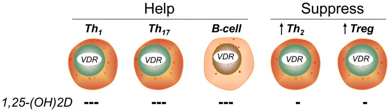
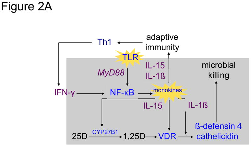
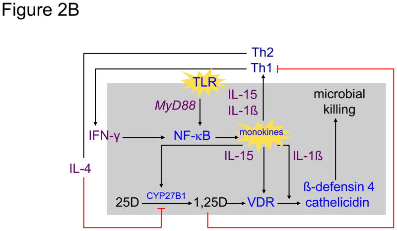
Like the vitamin D receptor (VDR), the CYP27B1-hydroxylase is expressed widely in human tissues. This expression profile establishes the potential for interaction of the VDR with the product of the CYP27B1, 1,25-dihydroxyvitamin D (1,25-(OH)(2)D), in either an intracrine or paracrine mode. This expansive expression profile also suggests that the local production and action of 1,25-(OH)(2)D to regulate VDR-directed gene expression may be similarly wide-ranging and distinct from what occurs in the kidney; the proximal renal tubular epithelial cell is the richest source of the CYP27B1 and the site for production of 1,25-(OH)(2)D destined to function as a hormone. Existence of the CYP27B1 at extrarenal sites has been widely documented, although the functional impact of the enzyme in these tissues has yet to be fully demonstrated. Two notable exceptions are the disease-activated macrophage (e.g., in sarcoidosis or tuberculosis) and the placenta. These two tissues are capable of generating enough 1,25-(OH)(2)D so as to be detectable in the general circulation. As such, this review will focus on CYP27B1 expression only at these two sites, theorizing that 1,25-(OH)(2)D production at these sites is for the purpose of local immunoregulatory function, not for controlling calcium balance in the host or the fetus.


Figures

Figure 1
Figure 1
Classes of lymphocytes designed to “help” (left; Th1, Th17 and immunoglobulin-producing B lymphocytes) and “suppress” (right; Th2 and Tregulatory [Treg] lymphocytes) the human immune response. When activated, all classes of lymphocytes express the vitamin D receptor (VDR). When exposed to 1,25-(OH)2D, the proliferation of each population is inhibited; the inhibition of proliferation is most profound in the “helper cell” classes, resulting in a net suppression of the adaptive immune response; the resultant relative increase in suppressor cell activity is indicated by the arrows.
Figure 2
Figure 2
Shown in the left panel 2A is a simplified schematic of the 25-hydroxyvitamin D (25D)-regulated innate immune response to Toll-like receptor (TLR) activation of the human macrophage in response to interaction with a pathogen-associated membrane pattern (PAMP) shed from Mycobacterium tuberculosis (mTB) [86]. Upon activation of the TLR 2/1 receptor by an mTB-specific PAMP, the activated receptor engages the intracellular coupling molecule MyD88 which, in turn, stimulates a kinase cascade ultimately leading to the nuclear localization of NF-κB and transcriptional activation of monokine genes, including IL-15 [102] and IL1-β [91]. In an autocrine fashion IL-15 promotes the expression of the CYP27B1 (1-hydroxlase) and vitamin D receptor (VDR) genes. In the presence of intracellularly-generated 1,25-dihydroxyvitamin D (1,25D), the ligand-bound VDR transactivates antimicrobial genes, cathelicidin and β-defensin 4, whose gene products, amplified under the autocrine actions of IL-1β, team with a 1,25D-directed autophagic response to promote microbial killing [96]. In a paracrine mode, IL-1β and IL-15 act to promote T helper-1 (Th1) cell generation of lymphokines, including interferon-gamma (IFN-γ), the most potent known stimulator of IL-15-driven CYP27B1 expression [103]. The net result is a feed-forward amplification loop to enhance microbial killing. The right panel 2B demonstrates interruption of the feed-forward loop by movement of 1,25D from the cell into the local inflammatory microenvironment when the CYP27B1-hydroxylating activity of the cell is robust. Escape of 1,25D permits it to interact with VDR-expressing helper (represented by Th1) and suppressor (represented Th2) cells [1]. Inhibition of “help” results in a decrease in IFN-γ production and downturn in macrophage 1,25D synthesis. Because the action of 1,25D to limit the proliferation and action of “suppressor” T cells is less strong, unopposed secretion of IL-4, a natural inhibitor of the CYP27B1, results in a further downturn in intracellular 1,25D synthesis and action.
Figure 2
Figure 2
Shown in the left panel 2A is a simplified schematic of the 25-hydroxyvitamin D (25D)-regulated innate immune response to Toll-like receptor (TLR) activation of the human macrophage in response to interaction with a pathogen-associated membrane pattern (PAMP) shed from Mycobacterium tuberculosis (mTB) [86]. Upon activation of the TLR 2/1 receptor by an mTB-specific PAMP, the activated receptor engages the intracellular coupling molecule MyD88 which, in turn, stimulates a kinase cascade ultimately leading to the nuclear localization of NF-κB and transcriptional activation of monokine genes, including IL-15 [102] and IL1-β [91]. In an autocrine fashion IL-15 promotes the expression of the CYP27B1 (1-hydroxlase) and vitamin D receptor (VDR) genes. In the presence of intracellularly-generated 1,25-dihydroxyvitamin D (1,25D), the ligand-bound VDR transactivates antimicrobial genes, cathelicidin and β-defensin 4, whose gene products, amplified under the autocrine actions of IL-1β, team with a 1,25D-directed autophagic response to promote microbial killing [96]. In a paracrine mode, IL-1β and IL-15 act to promote T helper-1 (Th1) cell generation of lymphokines, including interferon-gamma (IFN-γ), the most potent known stimulator of IL-15-driven CYP27B1 expression [103]. The net result is a feed-forward amplification loop to enhance microbial killing. The right panel 2B demonstrates interruption of the feed-forward loop by movement of 1,25D from the cell into the local inflammatory microenvironment when the CYP27B1-hydroxylating activity of the cell is robust. Escape of 1,25D permits it to interact with VDR-expressing helper (represented by Th1) and suppressor (represented Th2) cells [1]. Inhibition of “help” results in a decrease in IFN-γ production and downturn in macrophage 1,25D synthesis. Because the action of 1,25D to limit the proliferation and action of “suppressor” T cells is less strong, unopposed secretion of IL-4, a natural inhibitor of the CYP27B1, results in a further downturn in intracellular 1,25D synthesis and action.

References

    1. Hewison M. An update on vitamin D and human immunity. Clin Endocrinol (Oxf) 2011 - PubMed
    1. Hewison M, Burke F, Evans KN, Lammas DA, Sansom DM, Liu P, Modlin RL, Adams JS. Extra-renal 25-hydroxyvitamin D3-1alpha-hydroxylase in human health and disease. J Steroid Biochem Mol Biol. 2007;103:316–321. - PubMed
    1. Panda DK, Miao D, Tremblay ML, Sirois J, Farookhi R, Hendy GN, Goltzman D. Targeted ablation of the 25-hydroxyvitamin D 1alpha-hydroxylase enzyme: evidence for skeletal, reproductive, and immune dysfunction. Proc Natl Acad Sci U S A. 2001;98:7498–7503. - PMC - PubMed
    1. Vanhooke JL, Prahl JM, Kimmel-Jehan C, Mendelsohn M, Danielson EW, Healy KD, DeLuca HF. CYP27B1 null mice with LacZreporter gene display no 25-hydroxyvitamin D3-1alpha-hydroxylase promoter activity in the skin. Proceedings of the National Academy of Sciences of the United States of America. 2006;103:75–80. - PMC - PubMed
    1. St-Arnaud R, Dardenne O, Prud’homme J, Hacking SA, Glorieux FH. Conventional and tissue-specific inactivation of the 25-hydroxyvitamin D-1alpha-hydroxylase (CYP27B1) J Cell Biochem. 2003;88:245–251. - PubMed

Publication types

Substances