. 2013 Jan;50(1):200-8.
doi: 10.1177/0300985812439725. Epub 2012 Mar 23.

Novel genital alphapapillomaviruses in baboons (Papio hamadryas anubis) with cervical dysplasia

Affiliations

Novel genital alphapapillomaviruses in baboons (Papio hamadryas anubis) with cervical dysplasia

I L Bergin et al. Vet Pathol. 2013 Jan.

Abstract

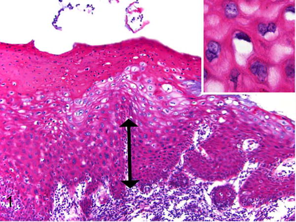
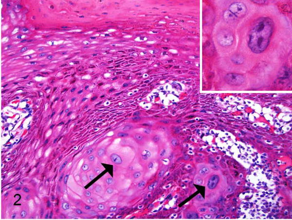
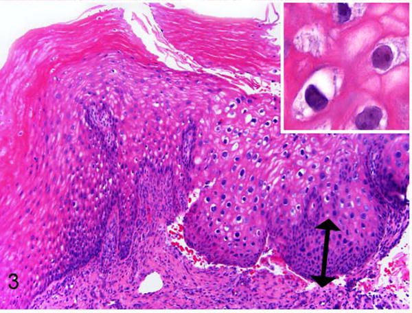
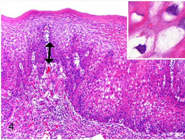
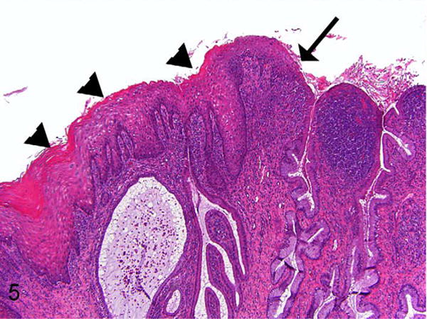
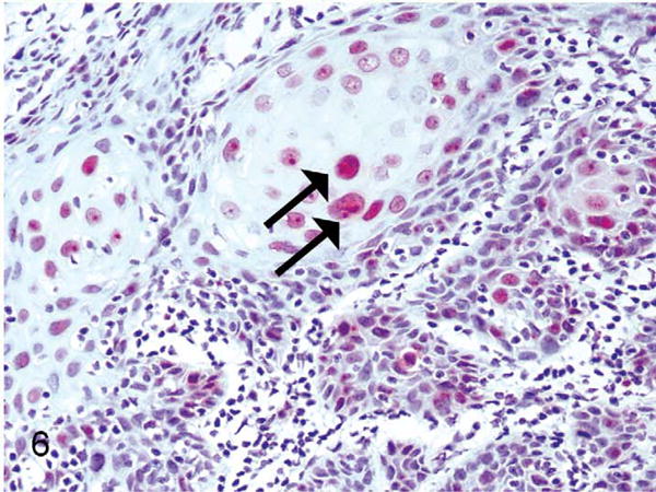
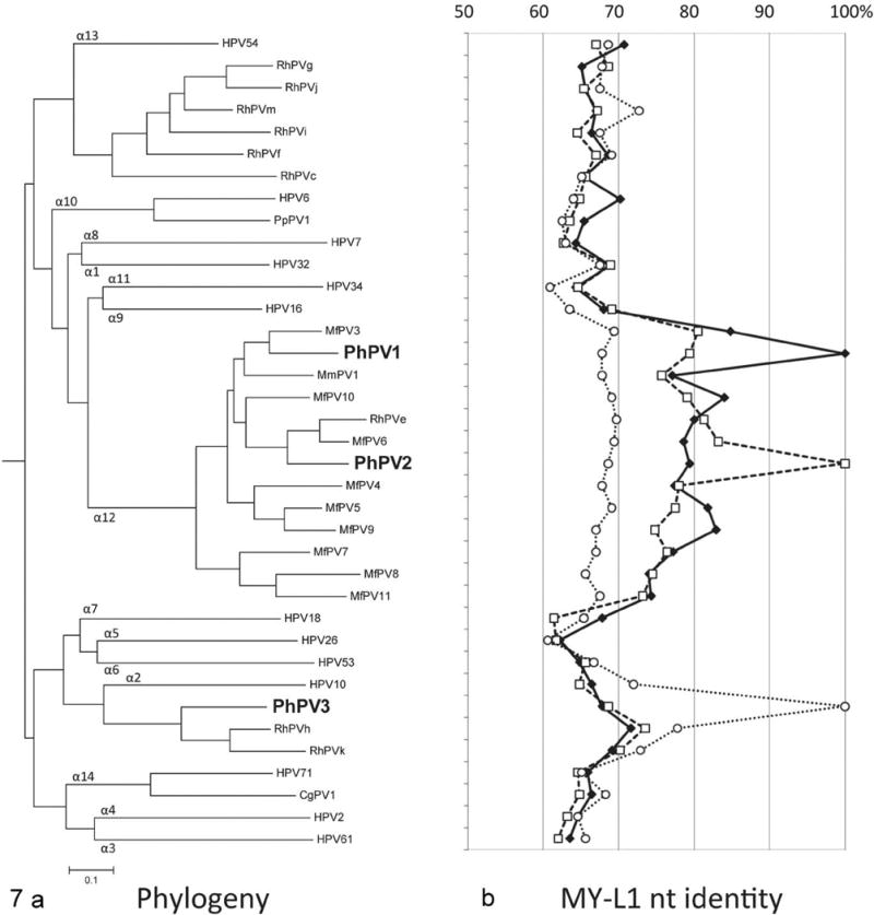
Genital Alphapapillomavirus (αPV) infections are one of the most common sexually transmitted human infections worldwide. Women infected with the highly oncogenic genital human papillomavirus (HPV) types 16 and 18 are at high risk for development of cervical cancer. Related oncogenic αPVs exist in rhesus and cynomolgus macaques. Here the authors identified 3 novel genital αPV types (PhPV1, PhPV2, PhPV3) by PCR in cervical samples from 6 of 15 (40%) wild-caught female Kenyan olive baboons (Papio hamadryas anubis). Eleven baboons had koilocytes in the cervix and vagina. Three baboons had dysplastic proliferative changes consistent with cervical squamous intraepithelial neoplasia (CIN). In 2 baboons with PCR-confirmed PhPV1, 1 had moderate (CIN2, n = 1) and 1 had low-grade (CIN1, n = 1) dysplasia. In 2 baboons with PCR-confirmed PhPV2, 1 had low-grade (CIN1, n = 1) dysplasia and the other had only koilocytes. Two baboons with PCR-confirmed PhPV3 had koilocytes only. PhPV1 and PhPV2 were closely related to oncogenic macaque and human αPVs. These findings suggest that αPV-infected baboons may be useful animal models for the pathogenesis, treatment, and prophylaxis of genital αPV neoplasia. Additionally, this discovery suggests that genital αPVs with oncogenic potential may infect a wider spectrum of non-human primate species than previously thought.


Conflict of interest statement

Declaration of Conflicting Interests: The authors declared no potential conflicts of interest with respect to the research, authorship, and/or publication of this article.

Figures

Figure 1
Figure 1
Exocervix, baboon No. 1. Squamous intraepithelial lesion assessed as moderate cervical intraepithelial neoplasia (CIN2). There is basal epithelial expansion (double arrow) affecting one-third to two-thirds of the mucosa. Koilocytes (epithelial cells with perinuclear halos and shrunken nuclei) and binucleated keratinocytes are present (inset). There is parakeratosis of the superficial epithelium, a feature commonly noted in baboons in this study. HE.
Figure 2
Figure 2
Exocervix, baboon No. 1. Squamous intraepithelial lesion from the same animal as in Figure 1, taken from a different exocervical site within the same section and also assessed as moderate dysplasia (CIN2). In addition to the basal epithelial expansion there is disorderly keratinocyte maturation with several large, hyperchromatic, and atypical cervical keratinocytes (arrows). Many are karyomegalic and there are frequent binucleated cells (inset). HE.
Figure 3
Figure 3
Exocervix, baboon No. 2. Area of epithelial hyperplasia and atypia assessed as CIN1 (low-grade). Basal epithelial expansion affects approximately one-third mucosal height (double arrow). This animal does not have the degree of atypia as in baboon No. 1, but there are koilocytes and individual karyomegalic cells present within the stratum spinosum (inset). HE.
Figure 4
Figure 4
Exocervix, baboon No. 3. Area of epithelial hyperplasia assessed as CIN1 (low-grade). Basal epithelial expansion affects < one-third mucosal height (double arrow). Normal polarity and maturation are maintained. There are numerous koilocytes (inset) with enlarged cells but minimal atypia (inset). HE.
Figure 5
Figure 5
Lower magnification of exocervix and squamocolumnar junction, baboon No. 2. This illustrates the proximity of parakeratotic cervical epithelium (arrowheads) to the squamocolumnar junction (arrow). This section is a recut from a deeper level of the block than what is shown in Figure 3. Parakeratosis of varying degrees was frequently observed at or near the squamocolumnar junction in multiple animals in the study. This figure also illustrates the cervical inflammation (predominantly lymphoplasmacytic) that was frequently present in the animals in this study. HE.
Figure 6
Figure 6
Exocervix, baboon No. 1. Suprabasilar nuclear immunostaining for the proliferation marker Ki67 (MIB1) (arrows) within a CIN2 lesion associated with PhPV1 infection.
Figure 7
Figure 7
Phylogeny of novel genital baboon papillomaviruses. a. Relationship of PhPV1, 2, and 3 to other genital Alphapapillomaviruses (αPVs). Maximum likelihood methods using the complete L1 nucleotide (capsid) sequence alignment of represented αPVs were used to infer the tree. The MY-L1 nucleotide sequences (partial capsid) were applied if the complete L1 ORF sequences were unavailable. Each αPV species group is indicated by number at branchpoints of the tree. PhPV1 and PhPV2 cluster with the α12 species group, as do the majority of oncogenic rhesus genital papillomaviruses. PhPV1, the type associated with high-grade CIN in one animal in this study, is most closely related to MfPV3, which is the most common oncogenic PV in macaques. HPV16 (highly oncogenic) is the most closely related human type, although it is a different species group (α9). PhPV3 clusters in a separate branch with two partially characterized non-oncogenic macaque types and (more distantly) HPV10, which is associated with epidermodysplasia verruciformis (benign epithelial proliferative disease) in humans. b. Percent nucleotide identity of PhPV1, 2, and 3 in comparison with other αPVs. Each line represents the percent nucleotide similarity, based on the MY-L1 nucleotide sequences, between one PhPV type and the other papillomaviruses on the tree. The solid line represents PhPV1, the long dashed line represents PhPV2, and the short dashed line represents PhPV3. For each point on the line, the percent nucleotide similarity can be read from the top of the graph. For example, PhPV1 (solid line) is 100% similar to itself (point value of the solid line adjacent to the position of PhPV1 on the phylogenetic tree) but only 85% similar to its nearest neighbor, MfPV3 (point value of the solid line adjacent to MfPV3). Abbreviations: RhPVe-k, m, rhesus macaque (Macaca mulatta) papillomaviruses; MfPV3-11, Macaca fascicularis PV 3-11; MmPV1, Macaca mulatta PV 1; PpPV1, Pan paniscus PV 1.

References

    1. Antonsson A, Hansson BG. Healthy skin of many animal species harbors papillomaviruses which are closely related to their human counterparts. J Virol. 2002;76:12537–12542. - PMC - PubMed
    1. Bell JD, Bergin IL, Harris LH, et al. The effects of a single cervical inoculation of Chlamydia trachomatis on the female reproductive tract of the baboon. J Infect Dis. 2011;204:1305–1312. - PMC - PubMed
    1. Bernard HU, Burk RD, Chen ZG, et al. Classification of papillomaviruses (PVs) based on 189 PV types and proposal of taxonomic amendments. Virology. 2010;401:70–79. - PMC - PubMed
    1. Burk RD, Ho GYF, Beardsley L, et al. Sexual behavior and partner characteristics are the predominant risk factors for genital human papillomavirus infection in young women. J Infecti Dis. 1996;174:679–689. - PubMed
    1. Campo MS. Animal models of papillomavirus pathogenesis. Virus Res. 2002;89:249–261. - PubMed

Publication types

MeSH terms