

Active emergence from propofol general anesthesia is induced by methylphenidate
- PMID: 22446983
- PMCID: PMC3339625
- DOI: 10.1097/ALN.0b013e3182518bfc
Active emergence from propofol general anesthesia is induced by methylphenidate
Abstract
Background: A recent study showed that methylphenidate induces emergence from isoflurane general anesthesia. Isoflurane and propofol are general anesthetics that may have distinct molecular mechanisms of action. The objective of this study was to test the hypothesis that methylphenidate actively induces emergence from propofol general anesthesia.
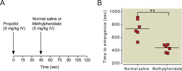
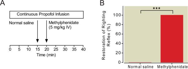
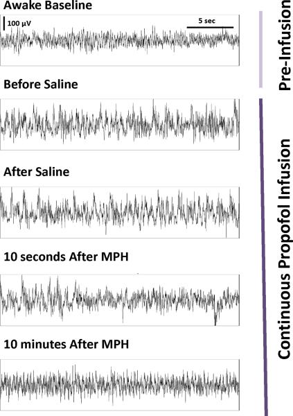
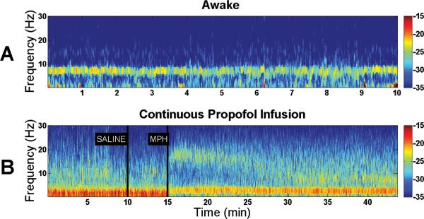
Methods: Using adult rats, the effect of methylphenidate on time to emergence after a single bolus of propofol was determined. The ability of methylphenidate to restore righting during a continuous target-controlled infusion (TCI) of propofol was also tested. In a separate group of rats, a TCI of propofol was established and spectral analysis was performed on electroencephalogram recordings taken before and after methylphenidate administration.
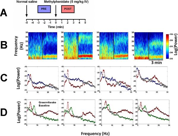
Results: Methylphenidate decreased median time to emergence after a single dose of propofol from 735 s (95% CI: 598-897 s, n = 6) to 448 s (95% CI: 371-495 s, n = 6). The difference was statistically significant (P = 0.0051). During continuous propofol anesthesia with a median final target plasma concentration of 4.0 μg/ml (95% CI: 3.2-4.6, n = 6), none of the rats exhibited purposeful movements after injection of normal saline. After methylphenidate, however, all six rats promptly exhibited arousal and had restoration of righting with a median time of 82 s (95% CI: 30-166 s). Spectral analysis of electroencephalogram data demonstrated a shift in peak power from δ (less than 4 Hz) to θ (4-8 Hz) and β (12-30 Hz) after administration of methylphenidate, indicating arousal in 4/4 rats.
Conclusions: Methylphenidate decreases time to emergence after a single dose of propofol, and induces emergence during continuous propofol anesthesia in rats. Further study is warranted to test the hypothesis that methylphenidate induces emergence from propofol general anesthesia in humans.
Figures
Comment in
-
From the edge of oblivion: the dance between intrinsic neuronal currents and neuronal connectivity.Anesthesiology. 2012 May;116(5):977-9. doi: 10.1097/ALN.0b013e3182518cc4. Anesthesiology. 2012. PMID: 22450471 No abstract available.
-
Should we use psychostimulant drugs to boost the emergence from general anesthesia?Anesthesiology. 2012 Dec;117(6):1393-4; author reply 1394-5. doi: 10.1097/ALN.0b013e318272d898. Anesthesiology. 2012. PMID: 23168433 No abstract available.
References
-
- Luo T, Leung LS. Involvement of tuberomamillary histaminergic neurons in isoflurane anesthesia. Anesthesiology. 2011;115:36–43. - PubMed
-
- Franks NP. General anaesthesia: From molecular targets to neuronal pathways of sleep and arousal. Nat Rev Neurosci. 2008;9:370–86. - PubMed
-
- Hudetz AG, Wood JD, Kampine JP. Cholinergic reversal of isoflurane anesthesia in rats as measured by cross-approximate entropy of the electroencephalogram. Anesthesiology. 2003;99:1125–31. - PubMed
-
- Alkire MT, McReynolds JR, Hahn EL, Trivedi AN. Thalamic microinjection of nicotine reverses sevoflurane-induced loss of righting reflex in the rat. Anesthesiology. 2007;107:264–72. - PubMed
-
- Meuret P, Backman SB, Bonhomme V, Plourde G, Fiset P. Physostigmine reverses propofol-induced unconsciousness and attenuation of the auditory steady state response and bispectral index in human volunteers. Anesthesiology. 2000;93:708–17. - PubMed
Publication types
MeSH terms
Substances
Grants and funding
LinkOut - more resources
Full Text Sources
Other Literature Sources
