. 2012 Jun;205(11):1709-18.
doi: 10.1093/infdis/jis254. Epub 2012 Mar 23.

Targeting pan-resistant bacteria with antibodies to a broadly conserved surface polysaccharide expressed during infection

Affiliations

Targeting pan-resistant bacteria with antibodies to a broadly conserved surface polysaccharide expressed during infection

David Skurnik et al. J Infect Dis. 2012 Jun.

Abstract

Background: New therapeutic targets for antibiotic-resistant bacterial pathogens are desperately needed. The bacterial surface polysaccharide poly-β-(1-6)-N-acetyl-glucosamine (PNAG) mediates biofilm formation by some bacterial species, and antibodies to PNAG can confer protective immunity. By analyzing sequenced genomes, we found that potentially multidrug-resistant bacterial species such as Klebsiella pneumoniae, Enterobacter cloacae, Stenotrophomonas maltophilia, and the Burkholderia cepacia complex (BCC) may be able to produce PNAG. Among patients with cystic fibrosis patients, highly antibiotic-resistant bacteria in the BCC have emerged as problematic pathogens, providing an impetus to study the potential of PNAG to be targeted for immunotherapy against pan-resistant bacterial pathogens.

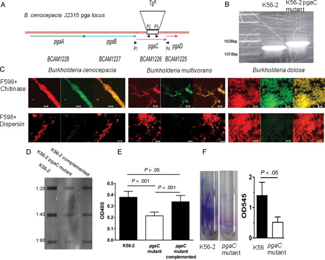
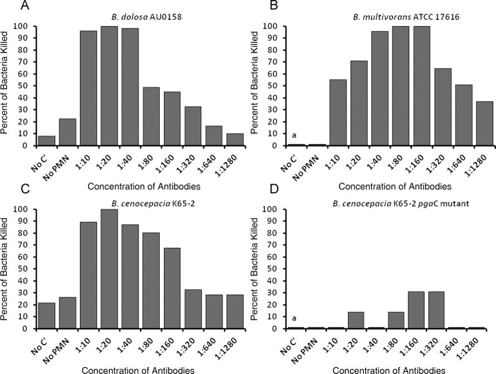
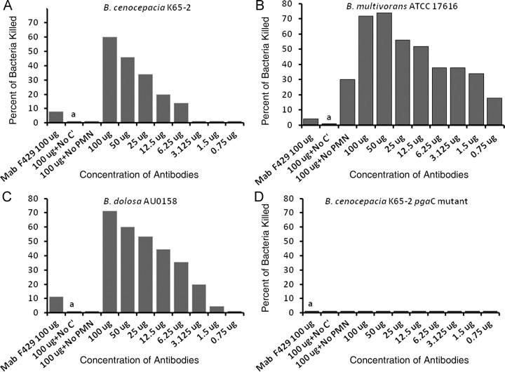
Methods: The presence of PNAG on BCC was assessed using a combination of bacterial genetics, microscopy, and immunochemical approaches. Antibodies to PNAG were tested using opsonophagocytic assays and for protective efficacy against lethal peritonitis in mice.

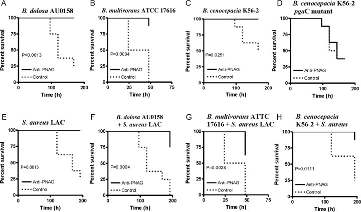
Results: PNAG is expressed in vitro and in vivo by the BCC, and cystic fibrosis patients infected by the BCC species B. dolosa mounted a PNAG-specific opsonophagocytic antibody response. Antisera to PNAG mediated opsonophagocytic killing of BCC and were protective against lethal BCC peritonitis even during coinfection with methicillin-resistant Staphylococcus aureus.

Conclusions: Our findings raise potential new therapeutic options against PNAG-producing bacteria, including even pan-resistant pathogens.


Figures

Figure 1.
Figure 1.
Production of poly-β-(1-6)-N-acetylglucosamine (PNAG) by the Burkholderia cepacia complex. A, Antibiotic susceptibility testing of a pan-resistant B. dolosa determined by disk diffusion and Etest. B, Confocal microscopic images of lung tissue sections from a CF patient who died of B. dolosa pneumonia and sepsis. A. Nuclear staining with TO-PRO-3 (blue). B. B. dolosa-specific mouse serum followed by a red secondary antibody. C. Rabbit antiserum to PNAG followed by a green secondary antibody. D. Phase contrast. E. Overlay of all channels. F. Negative control (normal rabbit serum) with TO-PRO-3 nuclear stain. Bar is 10 microns for A-E. C, B. dolosa pga locus. D, Results of a BLAST search using the sequence of the pgaC gene. E, Detection of PNAG production by confocal microscopy and specific enzymatic digestion using chitinase (top row) or dispersin B (bottom row) using red DNA stain (left panels), anti-PNAG human IgG1 mAb F598 with green secondary antibody (middle panels). Right panels are overlays of red and green channels. Bar is 10 microns.
Figure 2.
Figure 2.
Poly-β-(1-6)-N-acetylglucosamine (PNAG) expression by the Burkholderia cepacia complex is dependent on the pga locus and associated with biofilm production. A, Burkholderia cenocepacia pga locus in strain J2315. DNA sequence was obtained from the Sanger Center (http://www.sanger.ac.uk/Projects/B_cenocepacia/) and analyzed using the Gene Construction Kit (Textco Biotech). Open reading frames are marked, as are primer sites and the trimethoprim resistance (TpR) insert in pgaC. B, Polymerase chain reaction confirmation of the pgaC insertional mutant in B. cenocepacia K56-2. C, Confocal microscopic images of B. cenocepacia K56-2 and K56-2 pgaC mutant using red DNA stain (left panel), anti-PNAG human immunoglobulin G1 (IgG1) monoclonal antibody F598 [17], or a control anti–Pseudomonas aeruginosa alginate IgG1 antibody with green secondary antibody (middle panel). Right panel is overlay of red and green channels. Bar is 10 μm. D, Expression of PNAG as assessed by immunoblot of B. cenocepacia K56-2, K56-2 pgaC mutant, and complemented K56-2 pgaC mutant. E, Detection of PNAG production by enzyme-linked immunosorbent assay after 2 days of growth at 30°C, comparing B. cenocepacia K56-2, its pgaC mutant, and the complemented mutant. The values shown are the means ± SD of 9 wells of a representative experiment. P values by analysis of variance with Tukey multiple comparisons test. F, Biofilm assay using crystal violet staining of biofilm formed after 2 days of growth at room temperature comparing B. cenocepacia K56-2 and its pgaC mutant, visualized in left panel and quantified in right panel. Right panel bars represent means (n = 3), error bars SD. P value by unpaired t test.
Figure 3.
Figure 3.
Presence of opsonophagocytic killing activity specific to poly-β-(1-6)-N-acetylglucosamine (PNAG) in sera from cystic fibrosis (CF) patients chronically infected with Burkholderia dolosa. Opsonophagocytic killing (OPK) of Burkholderia cepacia complex by human sera from 13 CF patients chronically infected by B. dolosa. A: OPK of B. dolosa. B: OPK of B. multivorans. C: OPK of B. cenocepacia. D: OPK of B. cenocepacia pgaC mutant. Sera were adsorbed to remove antibodies not specific for PNAG. *P < .001 in comparison to any of the other groups by Kruskal-Wallis test with Dunn multiple comparison test. Abbreviations: C′, complement; PMN, polymorphonuclear leukocyte.
Figure 4.
Figure 4.
Animal antibodies to poly-β-(1-6)-N-acetylglucosamine (PNAG) mediate opsonic killing of Burkholderia cepacia complex strains. Opsonophagocytic killing of B. dolosa AU0158 K56-2 (A), B. multivorans ATCC17616 (B), B. cenocepacia K56-2 (C), and B. cenocepacia K56-2 pgaC mutant (D) by PNAG-specific goat antiserum. Bars represent mean percentage of killing relative to control containing normal goat serum. All SDs (not shown) were <15%. Assays done in duplicate. Abbreviations: C′, complement; PMN, polymorphonuclear leukocyte; a, absence of killing arbitrarily assigned as 1% killing.
Figure 5.
Figure 5.
Human monoclonal antibody (mAb) to poly-β-(1-6)-N-acetylglucosamine (PNAG) mediates opsonic killing of Burkholderia cepacia complex (BCC) isolates. Opsonophagocytic killing of BCC isolates (B. cenocepacia K56-2, 1a; B. multivorans ATCC17616, 1b; B. dolosa AU0158, 1c; B. cenocepacia K56-2 pgaC mutant, 1d) by human immunoglobulin G1 (IgG1) mAb F598 to PNAG. Control is human IgG1 mAb to Pseudomonas alginate (F429). Bars represent mean percentage of killing relative to the starting inoculum. All SDs (not shown) were <12%. Abbreviations: C′, complement; PMN, polymorphonuclear leukocyte; a, absence of killing arbitrarily assigned as 1% killing.
Figure 6.
Figure 6.
Antibodies to poly-β-(1-6)-N-acetylglucosamine (PNAG) protect against lethal peritonitis from PNAG-producing Burkholderia cepacia complex (BCC) even during coinfection with methicillin-resistant Staphylococcus aureus. Mice were passively immunized with PNAG-specific goat antiserum given intraperitoneally 24 hours before and 4 hours after intraperitoneal challenge with the indicated strains. Doses were 5 × 109 colony-forming units (CFUs) per mouse for BCC other than B. multivorans, which was 5 × 1010 CFU/mouse. S. aureus strain LAC was used at 109 CFU/mouse (E–H). Controls received normal goat serum. P values by log rank test, n = 8 mice per group.

References

    1. Kumarasamy KK, Toleman MA, Walsh TR, et al. Emergence of a new antibiotic resistance mechanism in India, Pakistan, and the UK: a molecular, biological, and epidemiological study. Lancet Infect Dis. 2010;10:597–602. - PMC - PubMed
    1. Waters V, Ratjen F. Multidrug-resistant organisms in cystic fibrosis: management and infection-control issues. Expert Rev Anti Infect Ther. 2006;4:807–19. - PubMed
    1. Aaron SD, Vandemheen KL, Ramotar K, et al. Infection with transmissible strains of Pseudomonas aeruginosa and clinical outcomes in adults with cystic fibrosis. JAMA. 2010;304:2145–53. - PubMed
    1. Dasenbrook EC, Checkley W, Merlo CA, Konstan MW, Lechtzin N, Boyle MP. Association between respiratory tract methicillin-resistant Staphylococcus aureus and survival in cystic fibrosis. JAMA. 2010;303:2386–92. - PubMed
    1. Kalish LA, Waltz DA, Dovey M, et al. Impact of Burkholderia dolosa on lung function and survival in cystic fibrosis. Am J Respir Crit Care Med. 2006;173:421–5. - PubMed

Publication types

MeSH terms

Substances