. 2012 Mar;46(2):171-8.
doi: 10.4103/0019-5413.93685.

Tuberculosis spine: Therapeutically refractory disease

Affiliations

Tuberculosis spine: Therapeutically refractory disease

Anil K Jain et al. Indian J Orthop. 2012 Mar.

Abstract

Background: India ranks second amongst the high-burden multi drug resistant tuberculosis (MDR-TB) countries, with an estimated incidence of 2.3% MDR-TB cases amongst the new cases and 17.2% amongst the previously treated cases. The diagnosis and treatment protocol for MDR-TB of the spine are not clearly established. We report outcome of a series of 15 cases of TB spine who were suspected to be therapeutically refractory cases (MDR-TB) on the basis of clinicoradiological failures of initial treatment.

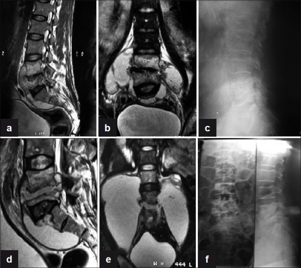
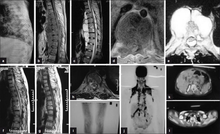
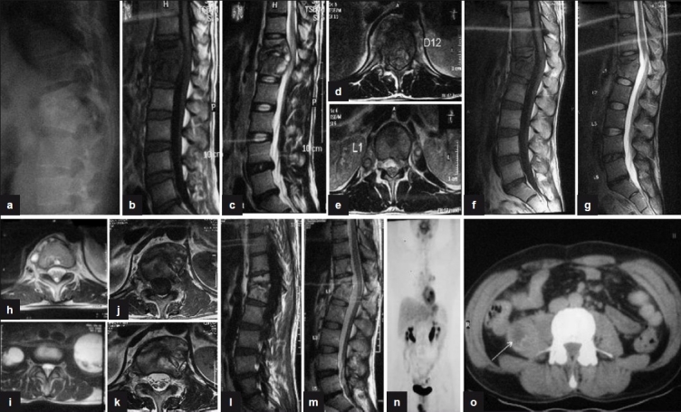
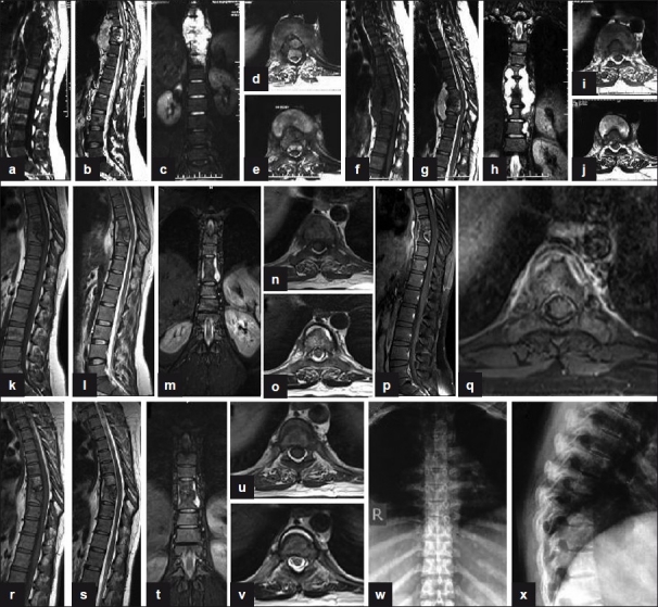
Materials and methods: Fifteen cases of TB spine from C2 to L5 spine were suspected to be the cases of MDR-TB (therapeutically refractory cases) on the basis of failures of adequate clinicoradiological healing response at 5 months or more on antitubercular treatment (ATT). None of the patient was immunocompromised. Thirteen out of 15 patients had tissue samples sent for histopathology, culture and sensitivity, smear, BACTEC, and polymerase chain reaction (PCR). All patients were put on second line ATT and followed up fortnightly with regular liver and kidney function tests, erythrocyte sedimentation rate (ESR), and plain X-ray. Healing was documented as subjective improvement of symptoms, reduction in ESR, and observations on contrast enhanced magnetic resonance imaging (MRI) such as resolution of marrow edema, fatty replacement of bone marrow and resolution of abscesses. Ambiguous MRI observations in a few patients were resolved on positron emission tomography (PET) scan. Patients were monitored continuously for 2 years after stopping ATT.

Results: We could demonstrate a positive culture in three cases. Two of them had multi drug resistance. We could achieve healing status in 13 out of 14 patients after starting second line drugs, one patient is still on treatment while other patient with no drug resistance is responding well on ATT.

Conclusions: The suspicion of therapeutically refractory case is of paramount importance. Once suspected, surgery to procure tissue for diagnosis and culture is to be undertaken. The demonstration of drug resistance on culture may not be achieved in all TB spine cases and empiric drug regimen for MDR-TB is to be started. We have achieved the healed status with immunomodulation and second line ATT. The length of treatment needs to be monitored with MRI and PET scan.

Keywords: MRI; TB spine; multi drug resistance; therapeutically refractory disease.


Conflict of interest statement

Conflict of Interest: None.

Figures

Figure 1
Figure 1
(a) Sagittal T2WI shows anterior subligamentous spread of large prevertebral collection with interosseous caseation seen in L4, L5 vertebra with endplate erosions and reduced disc height with discitis. (b) Coronal T2WI shows large paravertebral collections with interosseous caseation. (c) Plain X-ray lateral view of lumbosacral spine shows near complete obliteration of L4, L5 disc space with endplate erosions and loss of vertebral height at 3 months. (d, e) T2WI (Sagittal and coronal) show increase in the pre- and paravertebral collections with further loss of vertebral body height of L5 vertebra at 6 months. (f) Plain X-ray AP and lateral views of lumbosacral spine show sharpening of paradiscal cortical margins with better defined disc space and sclerosis of endplates with no paravertebral shadows on final healing
Figure 2
Figure 2
(a) Plain X-ray (pretreatment) lateral view shows reduction in disc space in D4, D5, D6, and in D9, D10, D11 with endplate erosions and fuzzy paradiscal margins. (b, c) Sagittal T1W and T2W (pretreatment) sections show paradiscal lesions with intraosseous caseation, preserved discs with discitis, anterior subligamentous spread of large prevertebral collection, anterior and posterior epidural spread of collection compressing the spinal cord. (d, e) Axial T1W and T2W sections show septate prevertebral collection, intraosseous caseation with large anterior and posterior epidural collections, and 80% canal encroachment. (f, g) Sagittal T1W and T2W (post-treatment) sections show patchy replacement of marrow by fat seen as bright T1 signal with near-complete resolution of collections and marrow edema. (j, k, l) Coronal and axial sections of PET scan show no FDG uptake in vertebral column at 18 months, suggestive of complete healing
Figure 3
Figure 3
(a) Plain X-ray lateral view shows anterior wedge collapse of D12 and L1 vetebra with complete obliteration of disc space. (b, c) Sagittal T1W and T2W sections show paradiscal lesions with intraosseous caseation, preserved discs with anterior subligamentous spread of prevertebral collection, and anterior epidural spread of collection compressing the spinal cord. (d, e) T2WI (axial) of D12 and L1 show septate prevertebral collection, intraosseous caseation with large anterior epidural collection, and 60% canal encroachment. (f, g) Sagittal T1WI and T2WI show persistent intraosseous caseation with decrease in epidural collection. (h, i) T2WI (axial) shows persistent septate collections with bilateral psoas collections at 18 months. (j, k) T1WI and T2WI (axial) show near complete resolution of paravertebral collections with residual marrow edema and no epidural extension at 24 months. (l, m) Sagittal T1WI and T2WI show fatty replacement of marrow in D12 and L1 vertebra seen as bright T1 signal with near complete resolution of collections and marrow edema at 30 months. (n, o) Coronal and axial PET scan shows persistent low grade activity in right psoas with no FDG uptake in the vertebra involved
Figure 4
Figure 4
(a, b) Sagittal T1W and T2W (preoperative) sections show paradiscal lesions with intraosseous caseation, preserved discs with discitis, anterior subligamentous spread of large prevertebral collection, anterior epidural spread of collection compressing the spinal cord. (c) Coronal T2W section shows large paravertebal collection. (d, e) Axial T1W and T2W sections show septate prevertebral collection, intraosseous caseation with large anterior epidural collections, and 80% canal encroachment. (f, g) Sagittal T1W and T2W (4-month post-op) sections show persistent collections and further destruction of D7, D8, and D9 vertebrae. (h) Coronal T2W section shows persistent large paravertebal collection. (i, j) Axial T1W and T2W sections show persistent septate loculated collections and anterior epidural extension. (k, l, m, n, o) MR sagittal, coronal, and axial (at 9 months of second line ATT) sections show significant resolution of collections with persistence of a thin rim of prevertebral collections with preserved discs and near-complete resolution of anterior epidural abscess. (p, q) Post-Gd-DTPA contrast sagittal and axial T1W sections show a thin rim of paravertebral abscess with complete resolution of anterior epidural abscess. (r, s, t, u, v) MR sagittal, coronal, and axial (at 14 months of second line ATT) sections show significant resolution of collections with patchy replacement of bone marrow by fat in D8, D9 vertebrae. A thin rim of anterior prevertebral collection is seen. (w, x) Plain X-ray AP and lateral views (at 18 months of second-line ATT) show sharpening and sclerosis of paradiscal margins with no significant paravertebral shadows suggestive of healing

References

    1. The World Health Organization/International Union Against Tuberculosis and Lung Disease (WHO/UNION) Global Project on Anti-Tuberculosis Drug Resistance Surveillance 2002–2007 Anti-Tuberculosis Drug Resistance In The World: Fourth Global Report
    1. Park K. 21st ed. Jabalpur, India: M/s Banarsidas Bhanot Publishers; 2011. Park textbook of preventive and social medicine; p. 164.
    1. Croften J, Mitchison D. Streptomycin resistence in pulmonary tuberculosis. Br Med J. 1948;2:1009–15. - PMC - PubMed
    1. Pyle MM. Relative numbers of resistant tubercle bacilli in sputum of patients before and during treatment with streptomycin. Proc Mayo Clin. 1947;22:465–73. - PubMed
    1. Tuli SM. Challenge of therapeutically refractory and multidrug resistant tuberculosis in orthopaedic practice. Indian J Orthop. 2002;36:211–3.

LinkOut - more resources