. 2012 Mar;46(2):191-9.
doi: 10.4103/0019-5413.93686.

Tuberculosis of hip in children: A retrospective analysis

Affiliations

Tuberculosis of hip in children: A retrospective analysis

Myung-Sang Moon et al. Indian J Orthop. 2012 Mar.

Abstract

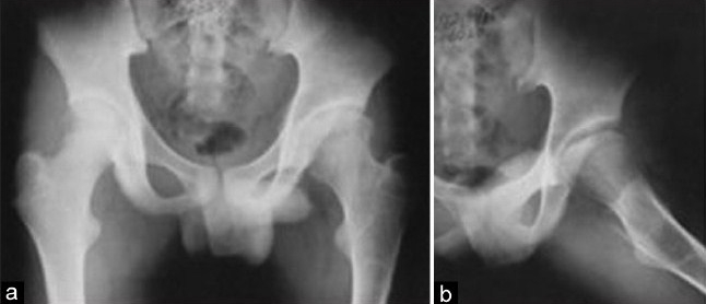
Background: Tuberculosis (TB) of hip constitutes nearly 15% of all cases of osteoarticular tuberculosis. We report a retrospective study carried out on 43 children with hip TB.

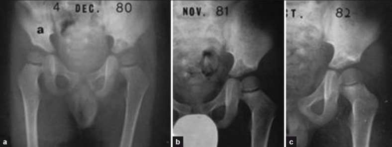
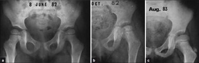
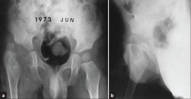
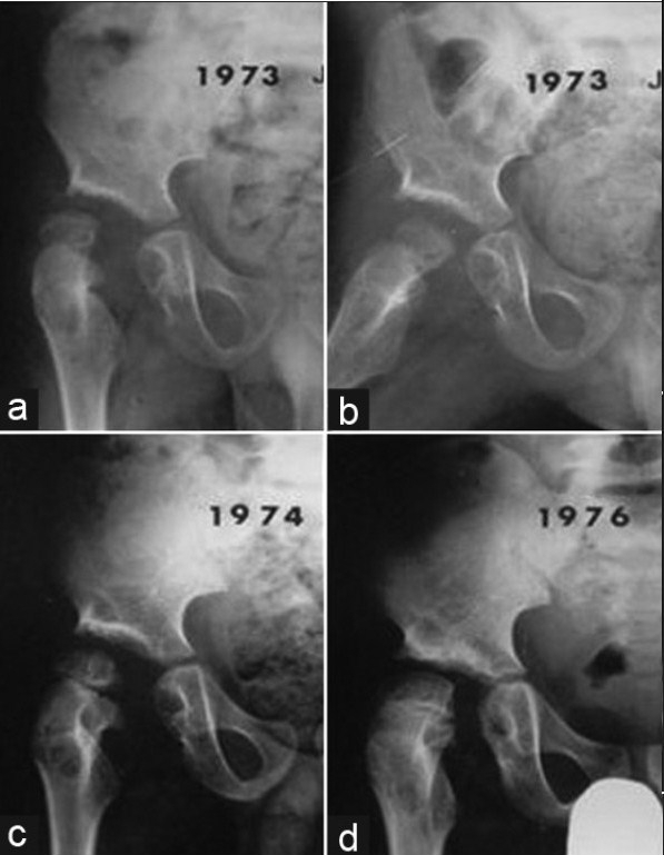
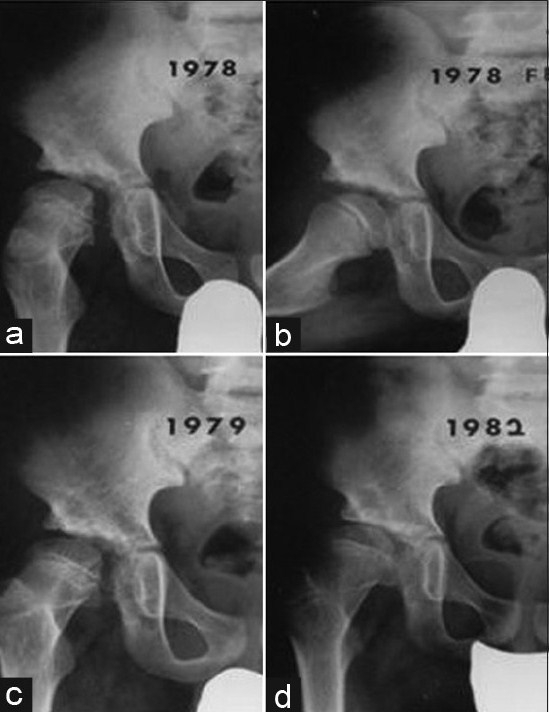
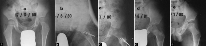
Materials and methods: Forty-three children of TB hip treated between 1971 and 2000 were analysed. Twenty-four children of the early series were treated with streptomycin (S), isoniazid (H) and PAS (Pa) for 18 months (3HPaS, 15 HPa), while 19 children in the later series were treated with isoniazid (H), rifampicin (R) and ethambutol (E) or pyrazinamide (Z) for 12 months [(12 RHE(Z)]. Five out of 18 children with radiologically normal appearing type hip TB were treated with chemotherapy alone and 38 children were subjected to surgery; simple synovectomy alone in 31 hips, joint debridement in six hips, and proximal femoral varisation osteotomy in one. After surgery hips were immobilized in cast for one to three months according to the severity of the disease and patients pain tolerance, and then were mobilized under leg traction in bed gradually till pain subsided completely.

Results: TB of hip healed with minimum sequelae in all children. In 18 Type one hip TB, normal hip (synovial form) anatomy was maintained, and in 25 patients with advanced lesions some defect in the femoral head and acetabulum was noticed, though painless good hip motion was maintained. Excellent to good results were obtained in 31 children (73.1%), fair in eight (18.6%), and poor in four (9.3%). In four patients with poor results, there was some residual morphological defect in the hip. None developed ankylosis of hip.

Conclusion: We achieved good outcome with minimum sequelae in this series. The management goal should be aimed not only to heal the disease but also to maintain a painless mobile hip and anatomical cephalocotyloid relationship until maturity, and retard the development of secondary osteoarthritis.

Keywords: Tuberculosis; hip; infective arthritis; osteoarticular.


Conflict of interest statement

Conflict of Interest: None.

Figures

Figure 1A
Figure 1A
Radiological types of tuberculosis of the hip, redrawn from Shanmugasundaram (1983)
Figure 1B
Figure 1B
Classification of tuberculosis of hip in children, based on the joint stability, cephalocotyloid morphology and its anatomical relation with Shanmugasundaran's classification (Moon, 1984)
Figure 2
Figure 2
X-ray pelvis with both hips anteroposterior view (a) and (L) hip joint (b, c) showing an example of normal hip type tuberculosis secondary to iliac acetabular lesion of left hip (a, b). Triple chemotherapy for 12 months and bed rest was given. The tuberculosis lesion had healed without joint damage and sequelae (c)
Figure 3
Figure 3
X-ray pelvis both hips (a) and (L) hip joint (b, c) anteroposterior view showing an example of Perthes type hip secondary to epiphysio-metaphyseal tuberculosis of the left proximal femur (a, b). After joint debridement, the child was immobilized with cast for 6 weeks under cover of triple chemotherapy (12 months). Tuberculosis healed without joint destruction, though residual coxa magna deformity was present (c)
Figure 4
Figure 4
X-ray pelvis both hips (a) and (L) hip joint (b, c, d) anteroposterior view showing an example of atrophic type hip due to tuberculosis of hip in a 11-year-old girl. There is slight joint space narrowing of left hip with slight pubes varus deformity of left pelvis (a). After synovectomy chemotherapy for 12 months was given (b, c, d). Tuberculosis healed (3 years followup) though atrophic head and neck are seen (d)
Figure 5
Figure 5
X-ray pelvis both hips (a) and (L) hip joint (b, c) anteroposterior view showing an example of relatively advanced tuberculosis of left hip in a 12-year-old boy (a). After joint debridement of left hip, hip was casted for 2 months under the cover of triple chemotherapy (b). However, there was progressive joint destruction. Therefore, chemotherapy was changed to 4 drug regimen. Finally mortar and pestle type of hip was seen (c). However the boy was free from pain, and his hip was clinically stable. He could enjoy almost normal life
Figure 6A
Figure 6A
X-ray pelvis both hips (a) and (R) hip joint (b) anteroposterior view showing an example of dislocating type of tuberculosis of hip in a 3-year-old boy. Multiple cystic lesions on epiphysis, metaphysis of the proximal femur, and medial aspect of the ischium, with an enlarged acetabulum (mortar type) destructive change are seen, but triradiate cartilage is well preserved
Figure 6B
Figure 6B
(a, b, c, d) followup radiograms (R) hip of same patient demonstrate the spontaneous gradual medial head migration and disappearance of the cystic bony lesions, though there is slight femoral head subluxation
Figure 6C
Figure 6C
(a, b, c, d) followup serial roentgenograms taken in February 1978, March 1979 and February 1982 still show some widening of the medial joint space with complete disappearance of the initial multiple cystic lesions on the epiphysis, metaphysis and ischium. Joint space narrowing and subchondral sclerosis of the right acetabular dome indicate the early osteoarthritic changes
Figure 7A
Figure 7A
X-ray pelvis with hips (a) and (L) hip joint anteroposterior view showing an example of travelling acetabulum in a 6-year-old boy. Hip was immobilized by cast for 2 months under cover of the triple antitubercular chemotherapy (a). Boy restored painless unlimited hip motion, though subluxation persisted on radiogram. Intertrochanteric femoral osteotomy was performed to treat the subluxed hip (b, c). Radiograms taken at postoperative one year and one year 6 months demonstrate the well seated femoral head in the acetabulum with slightly widened socket dome space (d, e)
Figure 7B
Figure 7B
Radiograms taken on postoperative 9 year (at age of 15 years) pelvis with both hips (a) and (L) hip (b) anteroposterior view demonstrates the good cephalocotyloid relationship, though some enlarged and flattened head with pubes varus deformity are seen

References

    1. Babhulkar S, Pande S. Tuberculosis of the hip. Clin Orthop Relat Res. 2002;398:93–9. - PubMed
    1. Campbell JA, Hoffman EB. Tuberculosis of the hip in children. J Bone Joint Surg Br. 1995;77:319–26. - PubMed
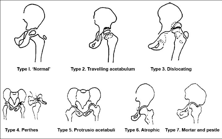
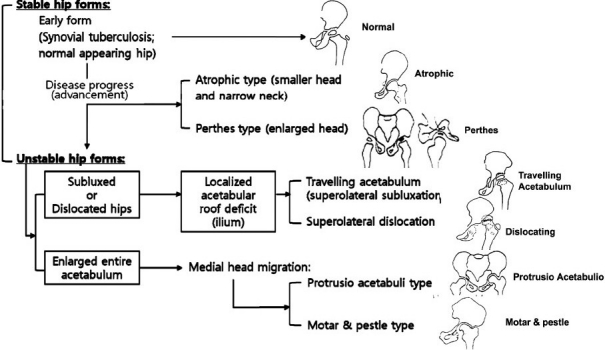
    1. Shanmugasundaram TK. A clinicoradiological classification of tuberculosis of the hip. In: Shanmugasundaram TK, editor. Current concepts in bone and joint tuberculosis. Madras, India: Proceedings of Combined Congress of International Bone and Joint Tuberculosis Club and the Indian Orthop Assoc; 1983. p. 60.
    1. Vora PH. Role of early surgery in management of tuberculosis of hip. In: Shanmugasundaram TK, editor. Current concept in bone and joint tuberculosis. Madras, India: Proceedings of Combined Congress of International Bone and Joint Tuberculosis Clue and the Indian Orthopedic Association; 1983. p. 60.
    1. Metha MT. Prognosis of tuberculous arthritis. In: Shanmugasundaram TK, editor. ibi-dem. Chennai: 1983. pp. 78–80.

LinkOut - more resources