

Recombinant vesicular stomatitis virus vaccine vectors expressing filovirus glycoproteins lack neurovirulence in nonhuman primates
- PMID: 22448291
- PMCID: PMC3308941
- DOI: 10.1371/journal.pntd.0001567
Recombinant vesicular stomatitis virus vaccine vectors expressing filovirus glycoproteins lack neurovirulence in nonhuman primates
Abstract
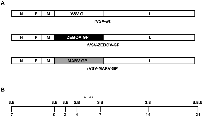
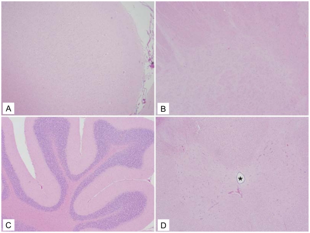
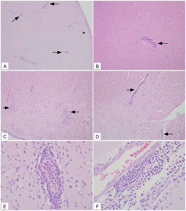
The filoviruses, Marburg virus and Ebola virus, cause severe hemorrhagic fever with high mortality in humans and nonhuman primates. Among the most promising filovirus vaccines under development is a system based on recombinant vesicular stomatitis virus (rVSV) that expresses an individual filovirus glycoprotein (GP) in place of the VSV glycoprotein (G). The main concern with all replication-competent vaccines, including the rVSV filovirus GP vectors, is their safety. To address this concern, we performed a neurovirulence study using 21 cynomolgus macaques where the vaccines were administered intrathalamically. Seven animals received a rVSV vector expressing the Zaire ebolavirus (ZEBOV) GP; seven animals received a rVSV vector expressing the Lake Victoria marburgvirus (MARV) GP; three animals received rVSV-wild type (wt) vector, and four animals received vehicle control. Two of three animals given rVSV-wt showed severe neurological symptoms whereas animals receiving vehicle control, rVSV-ZEBOV-GP, or rVSV-MARV-GP did not develop these symptoms. Histological analysis revealed major lesions in neural tissues of all three rVSV-wt animals; however, no significant lesions were observed in any animals from the filovirus vaccine or vehicle control groups. These data strongly suggest that rVSV filovirus GP vaccine vectors lack the neurovirulence properties associated with the rVSV-wt parent vector and support their further development as a vaccine platform for human use.
Conflict of interest statement
The authors have read the journal's policy and have the following conflicts: HF and TWG are named on patent applications for VSV-based vaccines for Ebola and Marburg viruses.
Figures
Similar articles
-
Single-Dose Trivalent VesiculoVax Vaccine Protects Macaques from Lethal Ebolavirus and Marburgvirus Challenge.J Virol. 2018 Jan 17;92(3):e01190-17. doi: 10.1128/JVI.01190-17. Print 2018 Feb 1. J Virol. 2018. PMID: 29142131 Free PMC article.
-
Recombinant vesicular stomatitis virus-based vaccines against Ebola and Marburg virus infections.J Infect Dis. 2011 Nov;204 Suppl 3(Suppl 3):S1075-81. doi: 10.1093/infdis/jir349. J Infect Dis. 2011. PMID: 21987744 Free PMC article. Review.
-
Single-injection vaccine protects nonhuman primates against infection with marburg virus and three species of ebola virus.J Virol. 2009 Jul;83(14):7296-304. doi: 10.1128/JVI.00561-09. Epub 2009 Apr 22. J Virol. 2009. PMID: 19386702 Free PMC article.
-
Vaccination With a Highly Attenuated Recombinant Vesicular Stomatitis Virus Vector Protects Against Challenge With a Lethal Dose of Ebola Virus.J Infect Dis. 2015 Oct 1;212 Suppl 2(Suppl 2):S443-51. doi: 10.1093/infdis/jiv316. Epub 2015 Jun 24. J Infect Dis. 2015. PMID: 26109675 Free PMC article.
-
Prospects for immunisation against Marburg and Ebola viruses.Rev Med Virol. 2010 Nov;20(6):344-57. doi: 10.1002/rmv.661. Rev Med Virol. 2010. PMID: 20658513 Free PMC article. Review.
Cited by
-
A Cloned Recombinant Vesicular Stomatitis Virus-Vectored Marburg Vaccine, PHV01, Protects Guinea Pigs from Lethal Marburg Virus Disease.Vaccines (Basel). 2022 Jun 23;10(7):1004. doi: 10.3390/vaccines10071004. Vaccines (Basel). 2022. PMID: 35891170 Free PMC article.
-
Ebola virus vaccines: an overview of current approaches.Expert Rev Vaccines. 2014 Apr;13(4):521-31. doi: 10.1586/14760584.2014.885841. Epub 2014 Feb 27. Expert Rev Vaccines. 2014. PMID: 24575870 Free PMC article. Review.
-
Single-dose live-attenuated Nipah virus vaccines confer complete protection by eliciting antibodies directed against surface glycoproteins.Vaccine. 2014 May 7;32(22):2637-44. doi: 10.1016/j.vaccine.2014.02.087. Epub 2014 Mar 12. Vaccine. 2014. PMID: 24631094 Free PMC article.
-
Live virus vaccines based on a vesicular stomatitis virus (VSV) backbone: Standardized template with key considerations for a risk/benefit assessment.Vaccine. 2016 Dec 12;34(51):6597-6609. doi: 10.1016/j.vaccine.2016.06.071. Epub 2016 Jul 6. Vaccine. 2016. PMID: 27395563 Free PMC article. Review.
-
Application of interferon modulators to overcome partial resistance of human ovarian cancers to VSV-GP oncolytic viral therapy.Mol Ther Oncolytics. 2016 Sep 28;3:16021. doi: 10.1038/mto.2016.21. eCollection 2016. Mol Ther Oncolytics. 2016. PMID: 27738655 Free PMC article.
References
-
- Feldmann H, Geisbert TW, Jahrling PB, Klenk HD, Netesov SV, et al., editors. Filoviridae. London: Elsevier/Academic Press; 2004. pp. 645–653.
-
- Sanchez A, Geisbert TW, Feldmann H, editors. (2006) Filoviridae: Marburg and Ebola Viruses. 5th ed. Philadelphia: Lippincott Williams & Wilkins; 2006. pp. 1409–1448.
-
- Hartman AL, Towner JS, Nichol ST. Ebola and marburg hemorrhagic fever. Clin Lab Med. 2010;30:161–177. - PubMed
-
- Hevey M, Negley D, Pushko P, Smith J, Schmaljohn A. Marburg virus vaccines based upon alphavirus replicons protect guinea pigs and nonhuman primates. Virology. 1998;251:28–37. - PubMed
Publication types
MeSH terms
Substances
LinkOut - more resources
Full Text Sources
Other Literature Sources
Medical
