. 2012 Mar;2(3):227-35.
doi: 10.1158/2159-8290.CD-11-0341. Epub 2012 Jan 16.

EGFR-mediated re-activation of MAPK signaling contributes to insensitivity of BRAF mutant colorectal cancers to RAF inhibition with vemurafenib

Affiliations

EGFR-mediated re-activation of MAPK signaling contributes to insensitivity of BRAF mutant colorectal cancers to RAF inhibition with vemurafenib

Ryan B Corcoran et al. Cancer Discov. 2012 Mar.

Abstract

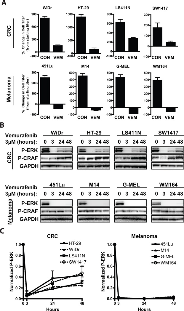
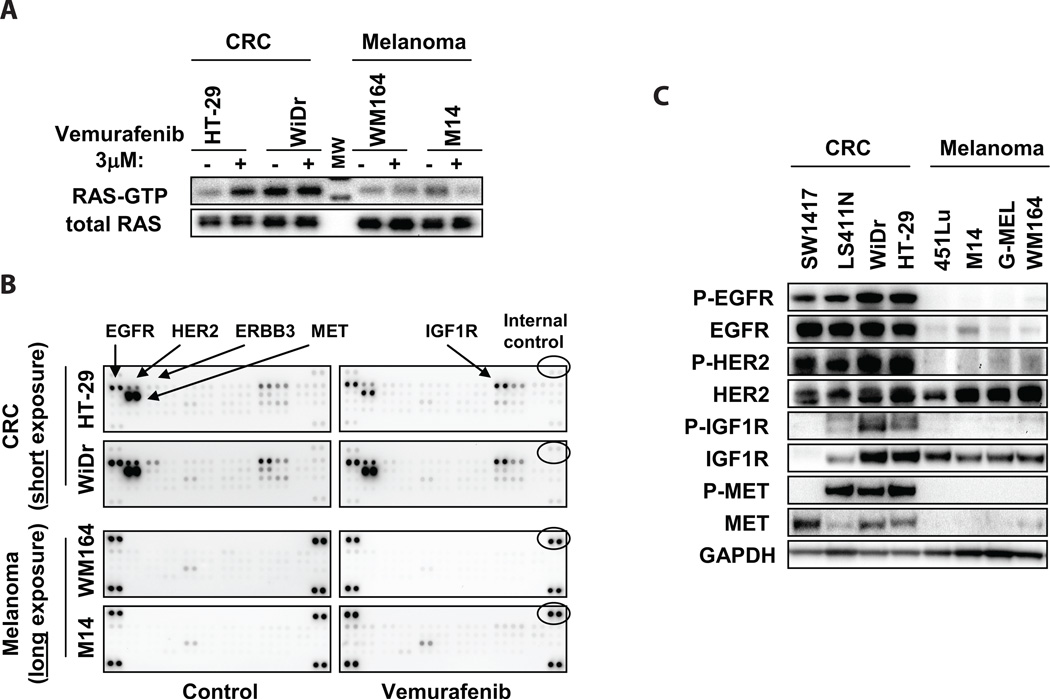
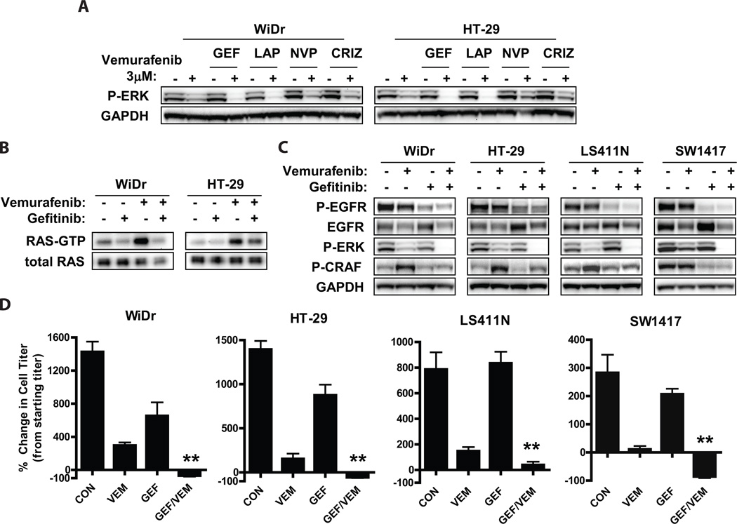
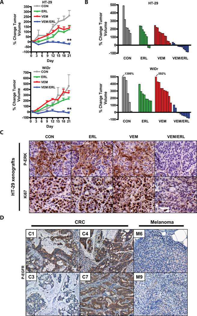
BRAF mutations occur in 10-15% of colorectal cancers (CRCs) and confer adverse outcome. While RAF inhibitors such as vemurafenib (PLX4032) have proven effective in BRAF mutant melanoma, they are surprisingly ineffective in BRAF mutant CRCs, and the reason for this disparity remains unclear. Compared to BRAF mutant melanoma cells, BRAF mutant CRC cells were less sensitive to vemurafenib, and P-ERK suppression was not sustained in response to treatment. Although transient inhibition of phospho-ERK by vemurafenib was observed in CRC, rapid ERK re-activation occurred through EGFR-mediated activation of RAS and CRAF. BRAF mutant CRCs expressed higher levels of phospho-EGFR than BRAF mutant melanomas, suggesting that CRCs are specifically poised for EGFR-mediated resistance. Combined RAF and EGFR inhibition blocked reactivation of MAPK signaling in BRAF mutant CRC cells and markedly improved efficacy in vitro and in vivo. These findings support evaluation of combined RAF and EGFR inhibition in BRAF mutant CRC patients.

Significance: BRAF valine 600 (V600) mutations occur in 10% to 15% of colorectal cancers, yet these tumors show a surprisingly low clinical response rate (~5%) to selective RAF inhibitors such as vemurafenib, which have produced dramatic response rates (60%–80%) in melanomas harboring the identical BRAF V600 mutation. We found that EGFR-mediated MAPK pathway reactivation leads to resistance to vemurafenib in BRAF-mutant colorectal cancers and that combined RAF and EGFR inhibition can lead to sustained MAPK pathway suppression and improved efficacy in vitro and in tumor xenografts.

Keywords: BRAF; EGFR; colorectal cancer; melanoma; vemurafenib.


Figures

Figure 1
Figure 1. Incomplete suppression of P-ERK in BRAF mutant CRCs is associated with decreased sensitivity to vemurafenib
(A.) BRAF mutant melanoma and CRC cell lines were treated with (VEM) or without (CON) 3µM vemurafenib for 72h, and viable cell titer was determined by Cell TiterGlo assay. Values represent the change in viable cell titer relative to the starting cell titer immediately prior to treatment. (B.) BRAF mutant cell lines from (A) were treated with 3µM vemurafenib for the indicated times, and lysates were probed with the indicated antibodies. (C.) Chemiluminescent quantifications of normalized P-ERK levels from western blots as in (B) are illustrated graphically. Values represent mean of three independent experiments.
Figure 2
Figure 2. Increased RTK activation in BRAF mutant CRC
(A.) Levels of active GTP-bound RAS were determined by RAS-GTP pulldown assay in the indicated cell lines treated with or without 3µM vemurafenib for 24h. (MW = molecular weight marker). (B.) Cells were treated with or without 3µM vemurafenib for 24h, and cell lysates were evaluated for levels of phosphorylated RTKs using phospho-RTK arrays. Short exposure is shown for BRAF mutant CRC cells, and long exposure is shown for BRAF mutant melanomas cells. Internal controls (indicated) allow comparison of absolute phospho-RTK levels between arrays. Key RTKs are indicated. (C.) Lysates from BRAF mutant CRC and melanoma cell lines were evaluated by western blot to determine total and phosphorylated protein levels of the RTKs identified in (B).
Figure 3
Figure 3. Combined inhibition of EGFR and RAF leads to sustained suppression of P-ERK and increased sensitivity in BRAF mutant CRC cells
(A.) BRAF mutant CRC cells were treated for 24h with or without 3µM vemurafenib in the presence or absence of gefitinib (GEF, 2 µM), lapatinib (LAP, 1µM), NVP-AEW541 (NVP, 1µM), or crizotinib (CRIZ, 1µM). Lysates were probed with the indicated antibodies. (B.) Cells were treated for 24h with the indicated inhibitors (vemurafenib 3µM, gefitinib 2µM) and levels of active GTP-bound RAS were determined by RAS-GTP pulldown assay. (C.) Cells were treated as indicated (vemurafenib 3µM, gefitinib 2µM) for 48h, and lysates were evaluated by western blot. (D.) BRAF mutant CRC cell lines were treated with 3µM vemurafenib or gefitinib 2µM, alone or in combination for 72h, and viable cell titer was determined by Cell TiterGlo assay. Values represent the change in viable cell titer relative to the starting cell titer immediately prior to treatment. Asterisks represent p <0.01.
Figure 4
Figure 4. Combined RAF and EGFR inhibition leads to improved in vivo efficacy in BRAF mutant CRC
(A.) BRAF mutant CRC xenografts derived from HT-29 and WiDr cells were treated with vehicle only (CON), vemurafenib only (VEM, 75mpk twice daily), erlotinib (ERL, 100mpk daily), or both inhibitors (VEM/ERL) in combination for 21d. Average percent change in tumor volume relative to initial tumor volume is shown. Error bars represent SEM. Asterisks represent p <0.001 for combined vemurafenib/erlotinib vs. all other treatment groups. (B.) Waterfall plots showing the percent change in volume (relative to initial tumor volume) for the individual tumors in each treatment group. (C.) Tumor tissue from HT-29 xenografts treated for 3d as indicated was evaluated by IHC for P-ERK and a marker of cell proliferation (Ki67). Tumors were harvested 4h after dosing on day 3. (D.) Levels of P-EGFR were assessed in human BRAF mutant CRCs and melanomas by IHC. Representative examples are shown. CRC cases with the lowest (C3) and highest (C7) P-EGFR levels are shown. 60% of BRAF mutant CRCs (n=10) exhibited high levels of P-EGFR, whereas only 18% of BRAF mutant melanomas (n=11) exhibited high levels of P-EGFR (p<0.05).

References

    1. Davies H, Bignell GR, Cox C, Stephens P, Edkins S, Clegg S, et al. Mutations of the BRAF gene in human cancer. Nature. 2002;417:949–954. - PubMed
    1. Montagut C, Settleman J. Targeting the RAF-MEK-ERK pathway in cancer therapy. Cancer Lett. 2009;283:125–134. - PubMed
    1. Yoon S, Seger R. The extracellular signal-regulated kinase: multiple substrates regulate diverse cellular functions. Growth Factors. 2006;24:21–44. - PubMed
    1. Hutchins G, Southward K, Handley K, Magill L, Beaumont C, Stahlschmidt J, et al. Value of mismatch repair, KRAS, and BRAF mutations in predicting recurrence and benefits from chemotherapy in colorectal cancer. J Clin Oncol. 2011;29:1261–1270. - PubMed
    1. Van Cutsem E, Kohne CH, Lang I, Folprecht G, Nowacki MP, Cascinu S, et al. Cetuximab plus irinotecan, fluorouracil, and leucovorin as first-line treatment for metastatic colorectal cancer: updated analysis of overall survival according to tumor KRAS and BRAF mutation status. J Clin Oncol. 2011;29:2011–2019. - PubMed

Publication types

MeSH terms