Review
. 2012 Sep;98(3):241-9.
doi: 10.1016/j.pneurobio.2012.02.004. Epub 2012 Mar 17.

High frequency oscillations in the intact brain

Affiliations
Review

High frequency oscillations in the intact brain

György Buzsáki et al. Prog Neurobiol. 2012 Sep.

Abstract

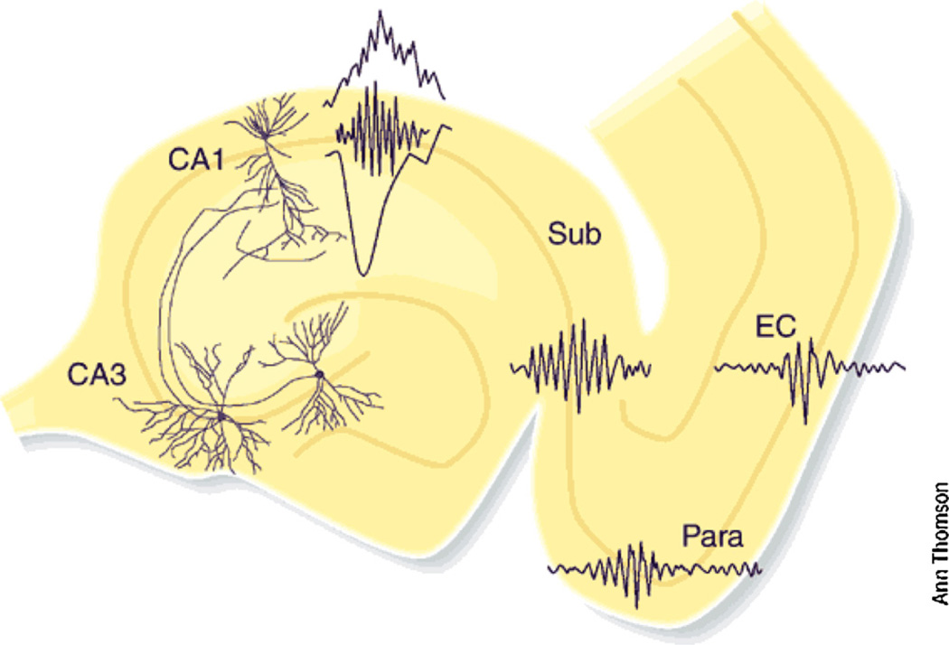
High frequency oscillations (HFOs) constitute a novel trend in neurophysiology that is fascinating neuroscientists in general, and epileptologists in particular. But what are HFOs? What is the frequency range of HFOs? Are there different types of HFOs, physiological and pathological? How are HFOs generated? Can HFOs represent temporal codes for cognitive processes? These questions are pressing and this symposium volume attempts to give constructive answers. As a prelude to this exciting discussion, we summarize the physiological high frequency patterns in the intact brain, concentrating mainly on hippocampal patterns, where the mechanisms of high frequency oscillations are perhaps best understood.


Figures

Fig. 1
Fig. 1
Self-organized burst of activity in the CA3 region of the hippocampus produces a sharp wave sink in the apical dendrites of CA1 pyramidal neurons and also discharge interneurons. The interactions between the discharging pyramidal cells and interneurons give rise to a short-lived fast oscillation (‘ripple’; 140–200 Hz), which can be detected as a field potential in the somatic layer. The strong CA1 population burst brings about strongly synchronized activity in the target populations of parahippocampal structures as well. These parahippocampal ripples are slower and less synchronous, compared to CA1 ripples. Reprinted from Buzsáki and Chrobak (2005).
Fig. 2
Fig. 2
Place cell sequences experienced during behavior are replayed in both the forward and reverse direction during awake SPW-Rs. Spike trains for 13 place fields on the track are shown before, during and after a single traversal. Sequences that occur during track running are reactivated during SPW-Rs both prior to and after the run, when the rat stays immobile. Forward replay (left inset, red box) occurs before traversal of the environment and reverse replay (right inset, blue box) after. The CA1 local field potential is shown on top and the animal’s velocity is shown below. Reprinted from Diba and Buzsáki (2007).
Fig. 3
Fig. 3
Gain and loss of excitation in different hippocampal-entorhinal regions during SPW-Rs. Population means of ripple-unit cross-correlograms in CA1, CA3 and dentate gyrus (DG) of the hippocampus and layers II, III and V of the entorhinal cortex (EC2, EC3, EC5). Principal cells and putative interneurons are shown in the left and middle columns, respectively. Peak of the ripple episode is time 0. Right column, Relative increase of neuronal discharge, normalized to the baseline (−200 to 200 ms) for both pyramidal cells (pyr, gray line) and interneurons (int, black line). The ratio between the relative peaks of pyramidal cells and interneurons is defined as ‘gain’. Note largest excitatory gain in CA1, flowed by CA3 and EC5. Gain is balanced in DG and EC2, whereas in EC3 inhibition dominates. Data from Mizuseki et al. (2009).
Fig. 4
Fig. 4
Spontaneously occurring fast ‘ripple’ oscillations (400–500 Hz) in the neocortex of the rat during high-voltage spindles. (A) Averaged high-voltage spindles and associated unit firing histograms from layers IV–VI. (B) Wide-band (a and a′; 1 Hz–5 kHz), filtered field (b and b′; 200–800 Hz), and filtered unit (c and c′; 0.5–5 kHz) traces from layers IV and V, respectively. (C) Averaged fast waves and corresponding unit histograms. The field ripples are filtered (200–800 Hz) derivatives of the wide-band signals recorded from 16 sites. Note the sudden phase-reversal of the oscillating waves (arrows) but locking of unit discharges (dashed lines). These phase reversed dipoles likely reflect synchronous discharge of layer 5 neurons in the vicinity of the recording electrode. Reprinted from Kandel and Buzsáki (1997).

References

    1. Anastassiou CA, Montgomery SM, Barahona M, Buzsáki G, Koch C. The effect of spatially inhomogeneous extracellular electric fields on neurons. J. Neurosci. 2010;30:1925–1936. - PMC - PubMed
    1. Battaglia FP, Sutherland GR, McNaughton BL. Hippocampal sharp wave bursts coincide with neocortical up-state transitions. Learn. Mem. 2004;11:697–704. - PMC - PubMed
    1. Behrens CJ, van den Boom LP, de Hoz L, Friedman A, Heinemann U. Induction of sharp wave-ripple complexes in vitro and reorganization of hippocampal networks. Nat. Neurosci. 2005;8:1560–1567. - PubMed
    1. Belluscio MA, Mizuseki K, Schmidt R, Kempter R, Buzsáki G. Cross-frequency phase-phase coupling between θ and γ oscillations in the hippocampus. J. Neurosci. 2012;32(2):423–435. - PMC - PubMed
    1. Both M, Bähner F, von Bohlen und Halbach O, Draguhn A. Propagation of specific network patterns through the mouse hippocampus. Hippocampus. 2008;18:899–908. - PubMed

Publication types