Review
. 2012:100:279-317.
doi: 10.1016/B978-0-12-387786-4.00009-9.

MicroRNAs in heart development

Affiliations
Review

MicroRNAs in heart development

Ramón A Espinoza-Lewis et al. Curr Top Dev Biol. 2012.

Abstract

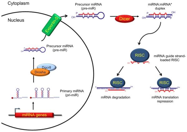
MicroRNAs (miRNAs) are a class of small noncoding RNAs of ~22nt in length which are involved in the regulation of gene expression at the posttranscriptional level by degrading their target mRNAs and/or inhibiting their translation. Expressed ubiquitously or in a tissue-specific manner, miRNAs are involved in the regulation of many biological processes such as cell proliferation, differentiation, apoptosis, and the maintenance of normal cellular physiology. Many miRNAs are expressed in embryonic, postnatal, and adult hearts. Aberrant expression or genetic deletion of miRNAs is associated with abnormal cardiac cell differentiation, disruption of heart development, and cardiac dysfunction. This chapter will summarize the history, biogenesis, and processing of miRNAs as well as their function in heart development, remodeling, and disease.


Figures

Figure 10.1
Figure 10.1
MicroRNA Biogenesis In the nucleus, a miRNA gene is transcribed into a long RNA molecule containing a single miRNA sequence or several miRNA sequences (polycistron or cluster), known as primary miRNA (pri-miRNA). Pri-miR-NAs are protected by the addition of a 5′-m7G cap (red ball) and a 3′ poly-A tail. Pre-miRNAs are processed by the microprocessor machinery composed of Drosha and the stabilizing protein Dgcr8, thus producing a precursor-miRNA molecule (pre-miRNA). Pre-miRNAs are transported out of the nucleus to the cytoplasm by the active trans-porter Exportin-5 and further processed by Dicer into a miRNA:miRNA* duplex. The RNA-induced silencing complex (RISC) is assembled and loaded with the miRNA guide strand due to the unwinding of the miRNA:miRNA* duplex. A loaded RISC will facilitate the recognition of mRNA targets leading to the mRNA degradation or the inhibition of translation.
Figure 10.2
Figure 10.2. Regulation and processing of the miR-1/miR-133 cluster
The miR-1/miR-133 clusters are present in different chromosomes (see text). Several transcription factor response elements are shown in the 5′upstream promoter region as well as in the “intergeneic” region. As shown, Nkx, CArG, E-box, and MEF-2 boxes represent recognition sites for Nkx2-5, SRF, Myogenin/MyoD, and MEF-2a binding, respectively. Further processing of the cluster results in the formation of two hairpin precursor miRNAs, miR-1 (in purple) and miR-133 (in red). Mature miRNA is represented in blue in both hairpins. Primary miRNAs are protected from early degradation by the addition of a 5′-m7G cap (red ball) and a 3′ poly-A tail.
Figure 10.3
Figure 10.3. miRNA function and integration in the regulatory networks to regulate cardiomyocyte differentiation and disease
Graphic representation of several regulatory networks targeted by miRNAs in cardiomyocyte differentiation from cardiac progenitor cells, cardiomyocytes, and their function in the regulation of cardiac hypertrophy. Also, the integration of miRNAs in such regulatory networks by targeting transcription factors, signaling molecules, and/or structural proteins is shown.
Figure 10.4
Figure 10.4. miRNA regulatory effects on cellular processes
Graphic representation of several miRNAs’ regulatory effects on two opposing cellular processes such as proliferation and apoptosis. It is worthy to note that one miRNA might be involved in both processes by targeting different molecules which, in turn, induce or inhibit such process.

References

    1. Abdellatif M. The role of microRNA-133 in cardiac hypertrophy uncovered. Circ. Res. 2010;106:16–18. - PMC - PubMed
    1. Alberts B, Bray D, Lewis J, Raff M, Roberts K, Watson J. Molecular Biology of the Cell. 3rd. Garland publishing; New York & London: 1994. pp. 72–73. 292–334.
    1. Ambros V, Horvitz HR. Heterochronic mutants of the Nematode Caenorhabditis elegans. Science. 1984;226:409–416. - PubMed
    1. Ambros V, Horvitz HR. The lin-14 locus of Caenorhabditis elegans controls the time of expression of specific postembryonic developmental events. Genes Dev. 1987;1:398–414. - PubMed
    1. Anderson P. Mutagenesis. In: Epstein F, Shakes C, editors. Caenorhabditis elegans: Modern Biological Analysis of an Organism. Academic press; California: 1995. pp. 31–54.

Publication types

LinkOut - more resources