

Molecular control of neuromuscular junction development
- PMID: 22450265
- PMCID: PMC3302983
- DOI: 10.1007/s13539-011-0041-7
Molecular control of neuromuscular junction development
Abstract
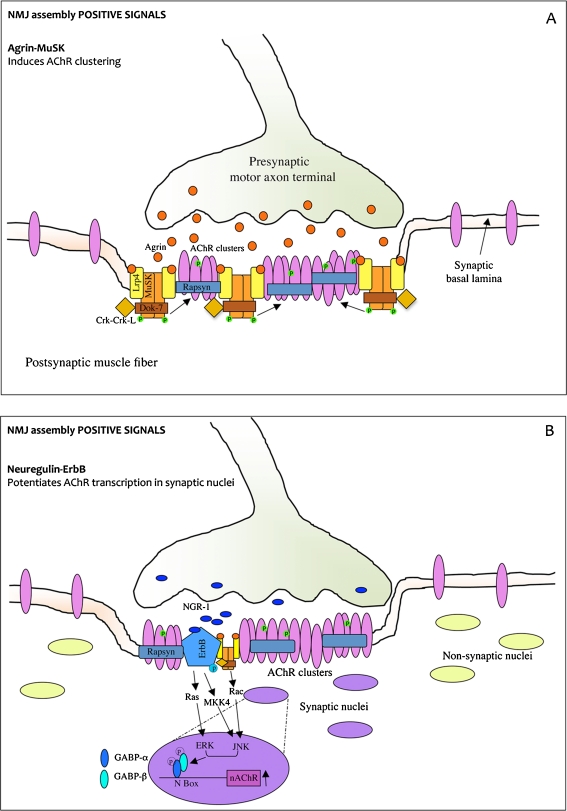
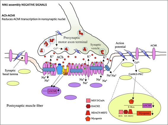
Skeletal muscle innervation is a multi-step process leading to the neuromuscular junction (NMJ) apparatus formation. The transmission of the signal from nerve to muscle occurs at the NMJ level. The molecular mechanism that orchestrates the organization and functioning of synapses is highly complex, and it has not been completely elucidated so far. Neuromuscular junctions are assembled on the muscle fibers at very precise locations called end plates (EP). Acetylcholine receptor (AChR) clusterization at the end plates is required for an accurate synaptic transmission. This review will focus on some mechanisms responsible for accomplishing the correct distribution of AChRs at the synapses. Recent evidences support the concept that a dual transcriptional control of AChR genes in subsynaptic and extrasynaptic nuclei is crucial for AChR clusterization. Moreover, new players have been discovered in the agrin-MuSK pathway, the master organizer of postsynaptical differentiation. Mutations in this pathway cause neuromuscular congenital disorders. Alterations of the postynaptic apparatus are also present in physiological conditions characterized by skeletal muscle wasting. Indeed, recent evidences demonstrate how NMJ misfunctioning has a crucial role at the onset of age-associated sarcopenia.
Figures
Similar articles
-
The synaptic muscle-specific kinase (MuSK) complex: new partners, new functions.Bioessays. 2005 Nov;27(11):1129-35. doi: 10.1002/bies.20305. Bioessays. 2005. PMID: 16237673 Review.
-
The role of nerve- versus muscle-derived factors in mammalian neuromuscular junction formation.J Neurosci. 2008 Mar 26;28(13):3333-40. doi: 10.1523/JNEUROSCI.5590-07.2008. J Neurosci. 2008. PMID: 18367600 Free PMC article.
-
[Genetic defects and disorders at the neuromuscular junction].Brain Nerve. 2011 Jul;63(7):669-78. Brain Nerve. 2011. PMID: 21747136 Review. Japanese.
-
The MuSK activator agrin has a separate role essential for postnatal maintenance of neuromuscular synapses.Proc Natl Acad Sci U S A. 2014 Nov 18;111(46):16556-61. doi: 10.1073/pnas.1408409111. Epub 2014 Nov 3. Proc Natl Acad Sci U S A. 2014. PMID: 25368159 Free PMC article.
-
Development of the neuromuscular junction.Cell Tissue Res. 2006 Nov;326(2):263-71. doi: 10.1007/s00441-006-0237-x. Epub 2006 Jul 4. Cell Tissue Res. 2006. PMID: 16819627 Review.
Cited by
-
Pre- and postsynaptic changes in the neuromuscular junction in dystrophic mice.Front Physiol. 2015 Sep 9;6:252. doi: 10.3389/fphys.2015.00252. eCollection 2015. Front Physiol. 2015. PMID: 26441672 Free PMC article.
-
Structural mechanisms of the agrin-LRP4-MuSK signaling pathway in neuromuscular junction differentiation.Cell Mol Life Sci. 2013 Sep;70(17):3077-88. doi: 10.1007/s00018-012-1209-9. Epub 2012 Nov 22. Cell Mol Life Sci. 2013. PMID: 23178848 Free PMC article. Review.
-
Bioreactor model of neuromuscular junction with electrical stimulation for pharmacological potency testing.Integr Biol (Camb). 2017 Dec 11;9(12):956-967. doi: 10.1039/c7ib00144d. Integr Biol (Camb). 2017. PMID: 29168874 Free PMC article.
-
Association of regular aerobic exercises and neuromuscular junction variants with incidence of frailty: an analysis of the Chinese Longitudinal Health and Longevity Survey.J Cachexia Sarcopenia Muscle. 2021 Apr;12(2):350-357. doi: 10.1002/jcsm.12658. Epub 2021 Feb 1. J Cachexia Sarcopenia Muscle. 2021. PMID: 33527771 Free PMC article.
-
Genetic deficiency of GABA differentially regulates respiratory and non-respiratory motor neuron development.PLoS One. 2013;8(2):e56257. doi: 10.1371/journal.pone.0056257. Epub 2013 Feb 15. PLoS One. 2013. PMID: 23457538 Free PMC article.
References
-
- Engel AG, Ohno K, Shen XM, Sine SM. Congenital myasthenic syndromes: multiple molecular targets at the neuromuscular junction. Ann N Y Acad Sci. 2003;998:138–160. - PubMed
-
- Hartzell HC, Fambrough DM. Acetycholine receptor production and incorporation into membranes of developing muscle fibers. Dev Biol. 1973;30:153–165. - PubMed
-
- Cohen SA, Fischbach GD. Clusters of acetylcholine receptors located at identified nerve-muscle synapses in vitro. Dev Biol. 1977;59:24–38. - PubMed
-
- Kummer TT, Misgeld T, Sanes JR. Assembly of the postsynaptic membrane at the neuromuscular junction: paradigm lost. Curr Opin Neurobiol. 2006;16:74–82. - PubMed
LinkOut - more resources
Full Text Sources
